 Movie
Movie Controller


+ Open data

- Basic information
| Entry | Database: PDB / ID: 7q1q | ||||||
|---|---|---|---|---|---|---|---|
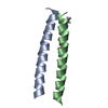
| Title | De novo designed homo-dimeric antiparallel helices Homomer-S | ||||||
 Components Components | Homomer-S | ||||||
 Keywords Keywords | DE NOVO PROTEIN / antiparallel homo-dimeric coiled-coil / de novo / protein design / associating peptides | ||||||
| Function / homology | ACETATE ION Function and homology information Function and homology information | ||||||
| Biological species | synthetic construct (others) | ||||||
| Method |  X-RAY DIFFRACTION / SYNCHROTRON / MOLECULAR REPLACEMENT / Resolution: 1 Å X-RAY DIFFRACTION / SYNCHROTRON / MOLECULAR REPLACEMENT / Resolution: 1 Å | ||||||
 Authors Authors | Shanmugaratnam, S. / Rhys, G.G. / Dawson, W.M. / Woolfson, D.N. / Hocker, B. | ||||||
| Funding support | European Union, 1items
| ||||||
 Citation Citation | Journal: Nat.Chem.Biol. / Year: 2022 Title: De novo designed peptides for cellular delivery and subcellular localisation. Authors: Rhys, G.G. / Cross, J.A. / Dawson, W.M. / Thompson, H.F. / Shanmugaratnam, S. / Savery, N.J. / Dodding, M.P. / Hocker, B. / Woolfson, D.N. | ||||||
| History |
|
- Structure visualization
| Structure viewer | Molecule: MolmilJmol/JSmol |
|---|
- Downloads & links
-Download
| PDBx/mmCIF format | 7q1q.cif.gz | 54.8 KB | Display | PDBx/mmCIF format |
|---|---|---|---|---|
| PDB format | pdb7q1q.ent.gz | 40.9 KB | Display | PDB format |
| PDBx/mmJSON format | 7q1q.json.gz | Tree view | PDBx/mmJSON format | |
| Others |  Other downloads Other downloads |
-Validation report
| Summary document | 7q1q_validation.pdf.gz | 429.9 KB | Display | wwPDB validaton report |
|---|---|---|---|---|
| Full document | 7q1q_full_validation.pdf.gz | 429.8 KB | Display | |
| Data in XML | 7q1q_validation.xml.gz | 5.5 KB | Display | |
| Data in CIF | 7q1q_validation.cif.gz | 6.8 KB | Display | |
| Arichive directory | https://data.pdbj.org/pub/pdb/validation_reports/q1/7q1qftp://data.pdbj.org/pub/pdb/validation_reports/q1/7q1q | HTTPS FTP |
-Related structure data


-Links
 PDBj
PDBj
- Assembly
| Deposited unit | 
| ||||||||
|---|---|---|---|---|---|---|---|---|---|
| 1 |
| ||||||||
| Unit cell |
|
-Components
| #1: Protein/peptide | Mass: 3332.809 Da / Num. of mol.: 2 / Source method: obtained synthetically / Source: (synth.) synthetic construct (others) #2: Chemical | ChemComp-ACT / |   Mass: 59.044 Da / Num. of mol.: 1 / Source method: obtained synthetically / Formula: C2H3O2 Mass: 59.044 Da / Num. of mol.: 1 / Source method: obtained synthetically / Formula: C2H3O2#3: Water | ChemComp-HOH / |  Mass: 18.015 Da / Num. of mol.: 63 / Source method: isolated from a natural source / Formula: H2O Mass: 18.015 Da / Num. of mol.: 63 / Source method: isolated from a natural source / Formula: H2OHas ligand of interest | N | Has protein modification | Y | |
|---|
-Experimental details
-Experiment
| Experiment | Method: X-RAY DIFFRACTION / Number of used crystals: 1 |
|---|
- Sample preparation
| Crystal | Density Matthews: 1.6 Å3/Da / Density % sol: 23.09 % |
|---|---|
| Crystal grow | Temperature: 293 K / Method: vapor diffusion, sitting drop / pH: 6.5 / Details: 1 M Acetate pH 6.5, 0.1 M imidazole |
-Data collection
| Diffraction | Mean temperature: 100 K / Serial crystal experiment: N |
|---|---|
| Diffraction source | Source: SYNCHROTRON / Site: BESSY  / Beamline: 14.2 / Wavelength: 0.9184 Å / Beamline: 14.2 / Wavelength: 0.9184 Å |
| Detector | Type: DECTRIS PILATUS3 2M / Detector: PIXEL / Date: Nov 7, 2019 |
| Radiation | Monochromator: DCM Si(111) / Protocol: SINGLE WAVELENGTH / Monochromatic (M) / Laue (L): M / Scattering type: x-ray |
| Radiation wavelength | Wavelength: 0.9184 Å / Relative weight: 1 |
| Reflection | Resolution: 1→17.08 Å / Num. obs: 20583 / % possible obs: 92.2 % / Redundancy: 3.5 % / Biso Wilson estimate: 10.6 Å2 / CC1/2: 0.999 / CC star: 1 / Rmerge(I) obs: 0.041 / Rpim(I) all: 0.026 / Rrim(I) all: 0.049 / Net I/σ(I): 12.69 |
| Reflection shell | Resolution: 1→1.04 Å / Redundancy: 3.4 % / Rmerge(I) obs: 1.351 / Mean I/σ(I) obs: 0.81 / Num. unique obs: 1960 / CC1/2: 0.451 / CC star: 0.788 / Rpim(I) all: 0.848 / Rrim(I) all: 1.6 / % possible all: 87.2 |
- Processing
| Software |
| ||||||||||||||||||||||||||||||||||||||||||||||||
|---|---|---|---|---|---|---|---|---|---|---|---|---|---|---|---|---|---|---|---|---|---|---|---|---|---|---|---|---|---|---|---|---|---|---|---|---|---|---|---|---|---|---|---|---|---|---|---|---|---|
| Refinement | Method to determine structure: MOLECULAR REPLACEMENT Starting model: Poly-Alanine coiled-coil Resolution: 1→17.08 Å / SU ML: 0.15 / Cross valid method: THROUGHOUT / Phase error: 28.65 / Stereochemistry target values: ML
| ||||||||||||||||||||||||||||||||||||||||||||||||
| Solvent computation | Shrinkage radii: 0.9 Å / VDW probe radii: 1.11 Å / Solvent model: FLAT BULK SOLVENT MODEL | ||||||||||||||||||||||||||||||||||||||||||||||||
| Displacement parameters | Biso max: 42.53 Å2 / Biso mean: 15.3 Å2 / Biso min: 7.32 Å2 | ||||||||||||||||||||||||||||||||||||||||||||||||
| Refinement step | Cycle: final / Resolution: 1→17.08 Å
| ||||||||||||||||||||||||||||||||||||||||||||||||
| LS refinement shell | Refine-ID: X-RAY DIFFRACTION / Rfactor Rfree error: 0
|
