 Movie
Movie Controller


+ Open data

- Basic information
| Entry | Database: PDB / ID: 7q0o | ||||||
|---|---|---|---|---|---|---|---|
| Title | E. coli NfsA | ||||||
 Components Components | Oxygen-insensitive NADPH nitroreductase | ||||||
 Keywords Keywords | OXIDOREDUCTASE / Oxygen-insensitive NADPH nitroreductase | ||||||
| Function / homology |  Function and homology information Function and homology informationNAD(P)H dehydrogenase (quinone) activity / Oxidoreductases / FMN binding / protein homodimerization activity / cytosol Similarity search - Function | ||||||
| Biological species |  | ||||||
| Method |  X-RAY DIFFRACTION / SYNCHROTRON / MOLECULAR REPLACEMENT / Resolution: 0.96 Å X-RAY DIFFRACTION / SYNCHROTRON / MOLECULAR REPLACEMENT / Resolution: 0.96 Å | ||||||
 Authors Authors | White, S.A. / Grainger, A. / Parr, R. / Day, M.A. / Jarrom, D. / Graziano, A. / Searle, P.F. / Hyde, E.I. | ||||||
| Funding support |  United Kingdom, 1items United Kingdom, 1items
| ||||||
 Citation Citation | Journal: Febs Lett. / Year: 2022 Title: The 3D-structure, kinetics and dynamics of the E. coli nitroreductase NfsA with NADP + provide glimpses of its catalytic mechanism. Authors: White, S.A. / Christofferson, A.J. / Grainger, A.I. / Day, M.A. / Jarrom, D. / Graziano, A.E. / Searle, P.F. / Hyde, E.I. | ||||||
| History |
|
- Structure visualization
| Structure viewer | Molecule: MolmilJmol/JSmol |
|---|
- Downloads & links
-Download
| PDBx/mmCIF format | 7q0o.cif.gz | 125.8 KB | Display | PDBx/mmCIF format |
|---|---|---|---|---|
| PDB format | pdb7q0o.ent.gz | 97.1 KB | Display | PDB format |
| PDBx/mmJSON format | 7q0o.json.gz | Tree view | PDBx/mmJSON format | |
| Others |  Other downloads Other downloads |
-Validation report
| Arichive directory | https://data.pdbj.org/pub/pdb/validation_reports/q0/7q0oftp://data.pdbj.org/pub/pdb/validation_reports/q0/7q0o | HTTPS FTP |
|---|
-Related structure data
| Related structure data |  7z0wC 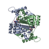 1f5vS S: Starting model for refinement C: citing same article ( |
|---|---|
| Similar structure data |
-Links
 PDBj
PDBj- Assembly
| Deposited unit | 
| ||||||||
|---|---|---|---|---|---|---|---|---|---|
| 1 | 
| ||||||||
| Unit cell |
| ||||||||
| Components on special symmetry positions |
|
-Components
| #1: Protein | Mass: 26832.664 Da / Num. of mol.: 1 Source method: isolated from a genetically manipulated source Source: (gene. exp.) |
|---|---|
| #2: Chemical | ChemComp-FMN /   Mass: 456.344 Da / Num. of mol.: 1 / Source method: obtained synthetically / Formula: C17H21N4O9P Mass: 456.344 Da / Num. of mol.: 1 / Source method: obtained synthetically / Formula: C17H21N4O9P |
| #3: Water | ChemComp-HOH /  Mass: 18.015 Da / Num. of mol.: 297 / Source method: isolated from a natural source / Formula: H2O Mass: 18.015 Da / Num. of mol.: 297 / Source method: isolated from a natural source / Formula: H2O |
| Has ligand of interest | Y |
-Experimental details
-Experiment
| Experiment | Method: X-RAY DIFFRACTION / Number of used crystals: 1 |
|---|
- Sample preparation
| Crystal | Density Matthews: 2.14 Å3/Da / Density % sol: 42.41 % |
|---|---|
| Crystal grow | Temperature: 291 K / Method: vapor diffusion, sitting drop / pH: 7.6 Details: 100 mM imidazole, pH 7.6, 20% PEG 6000, 200 mM Magnesium formate |
-Data collection
| Diffraction | Mean temperature: 100 K / Serial crystal experiment: N |
|---|---|
| Diffraction source | Source: SYNCHROTRON / Site: Diamond / Beamline: I04-1 / Wavelength: 0.9282 Å |
| Detector | Type: DECTRIS PILATUS 6M / Detector: PIXEL / Date: Feb 4, 2018 |
| Radiation | Protocol: SINGLE WAVELENGTH / Monochromatic (M) / Laue (L): M / Scattering type: x-ray |
| Radiation wavelength | Wavelength: 0.9282 Å / Relative weight: 1 |
| Reflection | Resolution: 0.96→41.28 Å / Num. obs: 132860 / % possible obs: 96.31 % / Redundancy: 1.8 % / Biso Wilson estimate: 10.82 Å2 / CC1/2: 0.997 / CC star: 0.999 / Rmerge(I) obs: 0.0217 / Rpim(I) all: 0.0217 / Rrim(I) all: 0.03069 / Net I/σ(I): 16.71 |
| Reflection shell | Resolution: 0.96→0.9943 Å / Redundancy: 1.4 % / Rmerge(I) obs: 0.2251 / Mean I/σ(I) obs: 2.12 / Num. unique obs: 10998 / CC1/2: 0.901 / CC star: 0.974 / Rpim(I) all: 0.2251 / Rrim(I) all: 0.3184 / % possible all: 79.86 |
- Processing
| Software |
| |||||||||||||||||||||||||||||||||||||||||||||||||||||||||||||||||
|---|---|---|---|---|---|---|---|---|---|---|---|---|---|---|---|---|---|---|---|---|---|---|---|---|---|---|---|---|---|---|---|---|---|---|---|---|---|---|---|---|---|---|---|---|---|---|---|---|---|---|---|---|---|---|---|---|---|---|---|---|---|---|---|---|---|---|
| Refinement | Method to determine structure: MOLECULAR REPLACEMENT Starting model: 1F5V Resolution: 0.96→41.28 Å / Cor.coef. Fo:Fc: 0.985 / Cor.coef. Fo:Fc free: 0.982 / SU B: 0.605 / SU ML: 0.014 / Cross valid method: THROUGHOUT / σ(F): 0 / ESU R: 0.016 / ESU R Free: 0.017 / Stereochemistry target values: MAXIMUM LIKELIHOOD Details: HYDROGENS HAVE BEEN ADDED IN THE RIDING POSITIONS U VALUES : REFINED INDIVIDUALLY
| |||||||||||||||||||||||||||||||||||||||||||||||||||||||||||||||||
| Solvent computation | Ion probe radii: 0.8 Å / Shrinkage radii: 0.8 Å / VDW probe radii: 1.2 Å / Solvent model: MASK | |||||||||||||||||||||||||||||||||||||||||||||||||||||||||||||||||
| Displacement parameters | Biso max: 229.52 Å2 / Biso mean: 19.105 Å2 / Biso min: 7.67 Å2
| |||||||||||||||||||||||||||||||||||||||||||||||||||||||||||||||||
| Refinement step | Cycle: final / Resolution: 0.96→41.28 Å
| |||||||||||||||||||||||||||||||||||||||||||||||||||||||||||||||||
| Refine LS restraints |
| |||||||||||||||||||||||||||||||||||||||||||||||||||||||||||||||||
| LS refinement shell | Resolution: 0.96→0.985 Å / Rfactor Rfree error: 0 / Total num. of bins used: 20
|
