 Movie
Movie Controller

[English] 日本語
 Yorodumi
- PDB-7pzg: Phocaeicola vulgatus sialic acid esterase at 1.44 Angstrom resolution -
+ Open data

- Basic information
| Entry | Database: PDB / ID: 7pzg | ||||||
|---|---|---|---|---|---|---|---|
| Title | Phocaeicola vulgatus sialic acid esterase at 1.44 Angstrom resolution | ||||||
 Components Components | Lysophospholipase L1 | ||||||
 Keywords Keywords | HYDROLASE / Serine hydrolase / Sialic acid acetyl esterase | ||||||
| Function / homology | : / SGNH hydrolase-type esterase domain / GDSL-like Lipase/Acylhydrolase family / SGNH hydrolase superfamily / phosphatidylcholine lysophospholipase A1 activity / Hydrolases; Acting on ester bonds / metal ion binding / MALONIC ACID / SGNH/GDSL hydrolase family protein Function and homology information Function and homology information | ||||||
| Biological species |  Phocaeicola vulgatus (bacteria) Phocaeicola vulgatus (bacteria) | ||||||
| Method |  X-RAY DIFFRACTION / SYNCHROTRON / MOLECULAR REPLACEMENT / Resolution: 1.44 Å X-RAY DIFFRACTION / SYNCHROTRON / MOLECULAR REPLACEMENT / Resolution: 1.44 Å | ||||||
 Authors Authors | Scott, H. / Armstrong, Z. / Davies, G.J. | ||||||
| Funding support |  United Kingdom, 1items United Kingdom, 1items
| ||||||
 Citation Citation | Journal: Acta Crystallogr D Struct Biol / Year: 2022 Title: The structure of Phocaeicola vulgatus sialic acid acetylesterase. Authors: Scott, H. / Davies, G.J. / Armstrong, Z. | ||||||
| History |
|
- Structure visualization
| Structure viewer | Molecule: MolmilJmol/JSmol |
|---|
- Downloads & links
-Download
| PDBx/mmCIF format | 7pzg.cif.gz | 340.8 KB | Display | PDBx/mmCIF format |
|---|---|---|---|---|
| PDB format | pdb7pzg.ent.gz | Display | PDB format | |
| PDBx/mmJSON format | 7pzg.json.gz | Tree view | PDBx/mmJSON format | |
| Others |  Other downloads Other downloads |
-Validation report
| Arichive directory | https://data.pdbj.org/pub/pdb/validation_reports/pz/7pzgftp://data.pdbj.org/pub/pdb/validation_reports/pz/7pzg | HTTPS FTP |
|---|
-Related structure data
| Related structure data | 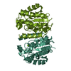 7pzhC  6njcS S: Starting model for refinement C: citing same article ( |
|---|---|
| Similar structure data |
-Links
 PDBj
PDBj
- Assembly
| Deposited unit | 
| ||||||||
|---|---|---|---|---|---|---|---|---|---|
| 1 | 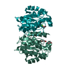
| ||||||||
| 2 | 
| ||||||||
| Unit cell |
|
-Components
| #1: Protein | Mass: 25227.924 Da / Num. of mol.: 4 Source method: isolated from a genetically manipulated source Source: (gene. exp.) Phocaeicola vulgatus (bacteria)Gene: DW869_04095, DWX04_04065, DXA04_10705, ERS852457_01148, ERS852509_00617, ERS852556_01195, F9001_09225, F9Z94_13025, GAS37_03660, GAX98_11820, GAY79_04625, GAY98_11740, GAZ06_07335, GAZ09_07170, ...Gene: DW869_04095, DWX04_04065, DXA04_10705, ERS852457_01148, ERS852509_00617, ERS852556_01195, F9001_09225, F9Z94_13025, GAS37_03660, GAX98_11820, GAY79_04625, GAY98_11740, GAZ06_07335, GAZ09_07170, GAZ65_18905, GAZ76_12675, GAZ90_18550, HKQ51_19175, HUV05_02570, SAMN04487923_100221 Production host: #2: Chemical | ChemComp-MG /   Mass: 24.305 Da / Num. of mol.: 4 / Source method: obtained synthetically / Formula: Mg Mass: 24.305 Da / Num. of mol.: 4 / Source method: obtained synthetically / Formula: Mg#3: Chemical | ChemComp-MLA /   Mass: 104.061 Da / Num. of mol.: 4 / Source method: obtained synthetically / Formula: C3H4O4 Mass: 104.061 Da / Num. of mol.: 4 / Source method: obtained synthetically / Formula: C3H4O4#4: Chemical | ChemComp-EDO /   Mass: 62.068 Da / Num. of mol.: 4 / Source method: obtained synthetically / Formula: C2H6O2 Mass: 62.068 Da / Num. of mol.: 4 / Source method: obtained synthetically / Formula: C2H6O2#5: Water | ChemComp-HOH / |  Mass: 18.015 Da / Num. of mol.: 696 / Source method: isolated from a natural source / Formula: H2O Mass: 18.015 Da / Num. of mol.: 696 / Source method: isolated from a natural source / Formula: H2OHas ligand of interest | N | |
|---|
-Experimental details
-Experiment
| Experiment | Method: X-RAY DIFFRACTION / Number of used crystals: 1 |
|---|
- Sample preparation
| Crystal | Density Matthews: 2.41 Å3/Da / Density % sol: 48.88 % |
|---|---|
| Crystal grow | Temperature: 293 K / Method: vapor diffusion, sitting drop / pH: 4.5 / Details: 18 % (w/v) PEG 3350, 200 mM sodium malonate pH 4.5 |
-Data collection
| Diffraction | Mean temperature: 100 K / Serial crystal experiment: N |
|---|---|
| Diffraction source | Source: SYNCHROTRON / Site: Diamond / Beamline: I03 / Wavelength: 0.97625 Å |
| Detector | Type: DECTRIS EIGER2 XE 16M / Detector: PIXEL / Date: Mar 12, 2021 |
| Radiation | Protocol: SINGLE WAVELENGTH / Monochromatic (M) / Laue (L): M / Scattering type: x-ray |
| Radiation wavelength | Wavelength: 0.97625 Å / Relative weight: 1 |
| Reflection | Resolution: 1.44→70.434 Å / Num. obs: 168248 / % possible obs: 97.7 % / Redundancy: 6.8 % / CC1/2: 1 / Net I/σ(I): 18.3 |
| Reflection shell | Resolution: 1.44→1.46 Å / Num. unique obs: 7900 / CC1/2: 0.712 |
- Processing
| Software |
| |||||||||||||||||||||||||||||||||||||||||||||||||||||||||||||||||||||||||||||||||||||||||||||||||||||||||||||||||||||||||||||||||||||||||||||||||||||||||||
|---|---|---|---|---|---|---|---|---|---|---|---|---|---|---|---|---|---|---|---|---|---|---|---|---|---|---|---|---|---|---|---|---|---|---|---|---|---|---|---|---|---|---|---|---|---|---|---|---|---|---|---|---|---|---|---|---|---|---|---|---|---|---|---|---|---|---|---|---|---|---|---|---|---|---|---|---|---|---|---|---|---|---|---|---|---|---|---|---|---|---|---|---|---|---|---|---|---|---|---|---|---|---|---|---|---|---|---|---|---|---|---|---|---|---|---|---|---|---|---|---|---|---|---|---|---|---|---|---|---|---|---|---|---|---|---|---|---|---|---|---|---|---|---|---|---|---|---|---|---|---|---|---|---|---|---|---|
| Refinement | Method to determine structure: MOLECULAR REPLACEMENT Starting model: 6njc Resolution: 1.44→70.434 Å / Cor.coef. Fo:Fc: 0.971 / Cor.coef. Fo:Fc free: 0.961 / WRfactor Rfree: 0.202 / WRfactor Rwork: 0.172 / SU B: 1.156 / SU ML: 0.044 / Average fsc free: 0.9248 / Average fsc work: 0.9304 / Cross valid method: FREE R-VALUE / ESU R: 0.061 / ESU R Free: 0.063 Details: Hydrogens have been added in their riding positions
| |||||||||||||||||||||||||||||||||||||||||||||||||||||||||||||||||||||||||||||||||||||||||||||||||||||||||||||||||||||||||||||||||||||||||||||||||||||||||||
| Solvent computation | Ion probe radii: 0.8 Å / Shrinkage radii: 0.8 Å / VDW probe radii: 1.2 Å / Solvent model: MASK BULK SOLVENT | |||||||||||||||||||||||||||||||||||||||||||||||||||||||||||||||||||||||||||||||||||||||||||||||||||||||||||||||||||||||||||||||||||||||||||||||||||||||||||
| Displacement parameters | Biso mean: 22.426 Å2
| |||||||||||||||||||||||||||||||||||||||||||||||||||||||||||||||||||||||||||||||||||||||||||||||||||||||||||||||||||||||||||||||||||||||||||||||||||||||||||
| Refinement step | Cycle: LAST / Resolution: 1.44→70.434 Å
| |||||||||||||||||||||||||||||||||||||||||||||||||||||||||||||||||||||||||||||||||||||||||||||||||||||||||||||||||||||||||||||||||||||||||||||||||||||||||||
| Refine LS restraints |
| |||||||||||||||||||||||||||||||||||||||||||||||||||||||||||||||||||||||||||||||||||||||||||||||||||||||||||||||||||||||||||||||||||||||||||||||||||||||||||
| LS refinement shell |
|
