 Movie
Movie Controller


+ Open data

- Basic information
| Entry | Database: PDB / ID: 7pud | ||||||
|---|---|---|---|---|---|---|---|
| Title | Bryoporin - actinoporin from moss Physcomitrium patens | ||||||
 Components Components | Bryoporin | ||||||
 Keywords Keywords | PLANT PROTEIN / Actinoporin / Membrane-binding / pore-formation / moss | ||||||
| Function / homology |  Function and homology information Function and homology informationpore complex assembly / cytolysis in another organism / pore complex / monoatomic cation transport / channel activity Similarity search - Function | ||||||
| Biological species |  Physcomitrium patens (plant) Physcomitrium patens (plant) | ||||||
| Method |  X-RAY DIFFRACTION / SYNCHROTRON / MOLECULAR REPLACEMENT / Resolution: 1.25 Å X-RAY DIFFRACTION / SYNCHROTRON / MOLECULAR REPLACEMENT / Resolution: 1.25 Å | ||||||
 Authors Authors | Solinc, G. / Anderluh, G. / Podobnik, M. | ||||||
| Funding support |  Slovenia, 1items Slovenia, 1items
| ||||||
 Citation Citation | Journal: J.Biol.Chem. / Year: 2022 Title: Pore-forming moss protein bryoporin is structurally and mechanistically related to actinoporins from evolutionarily distant cnidarians. Authors: Solinc, G. / Svigelj, T. / Omersa, N. / Snoj, T. / Pirc, K. / Znidarsic, N. / Yamaji-Hasegawa, A. / Kobayashi, T. / Anderluh, G. / Podobnik, M. | ||||||
| History |
|
- Structure visualization
| Structure viewer | Molecule: MolmilJmol/JSmol |
|---|
- Downloads & links
-Download
| PDBx/mmCIF format | 7pud.cif.gz | 66.1 KB | Display | PDBx/mmCIF format |
|---|---|---|---|---|
| PDB format | pdb7pud.ent.gz | 39 KB | Display | PDB format |
| PDBx/mmJSON format | 7pud.json.gz | Tree view | PDBx/mmJSON format | |
| Others |  Other downloads Other downloads |
-Validation report
| Arichive directory | https://data.pdbj.org/pub/pdb/validation_reports/pu/7pudftp://data.pdbj.org/pub/pdb/validation_reports/pu/7pud | HTTPS FTP |
|---|
-Related structure data
| Related structure data | 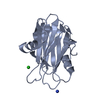 3vwiS S: Starting model for refinement |
|---|---|
| Similar structure data |
-Links
 PDBj
PDBj- Assembly
| Deposited unit | 
| ||||||||||||
|---|---|---|---|---|---|---|---|---|---|---|---|---|---|
| 1 |
| ||||||||||||
| Unit cell |
|
-Components
| #1: Protein | Mass: 21422.822 Da / Num. of mol.: 1 Source method: isolated from a genetically manipulated source Source: (gene. exp.) Physcomitrium patens (plant) / Gene: PHYPADRAFT_61094 / Plasmid: pET24a / Production host:  | ||||||
|---|---|---|---|---|---|---|---|
| #2: Chemical |   Mass: 118.174 Da / Num. of mol.: 2 / Source method: obtained synthetically / Formula: C6H14O2 / Comment: precipitant*YM Mass: 118.174 Da / Num. of mol.: 2 / Source method: obtained synthetically / Formula: C6H14O2 / Comment: precipitant*YM#3: Chemical | ChemComp-SO4 /   Mass: 96.063 Da / Num. of mol.: 8 / Source method: obtained synthetically / Formula: SO4 Mass: 96.063 Da / Num. of mol.: 8 / Source method: obtained synthetically / Formula: SO4#4: Water | ChemComp-HOH / |  Mass: 18.015 Da / Num. of mol.: 182 / Source method: isolated from a natural source / Formula: H2O Mass: 18.015 Da / Num. of mol.: 182 / Source method: isolated from a natural source / Formula: H2OHas ligand of interest | N | |
-Experimental details
-Experiment
| Experiment | Method: X-RAY DIFFRACTION / Number of used crystals: 1 |
|---|
- Sample preparation
| Crystal | Density Matthews: 1.97 Å3/Da / Density % sol: 37.6 % |
|---|---|
| Crystal grow | Temperature: 293.15 K / Method: vapor diffusion, hanging drop / Details: ammonium sulphate, NaCl |
-Data collection
| Diffraction | Mean temperature: 100 K / Serial crystal experiment: N |
|---|---|
| Diffraction source | Source: SYNCHROTRON / Site: ELETTRA  / Beamline: 5.2R / Wavelength: 1 Å / Beamline: 5.2R / Wavelength: 1 Å |
| Detector | Type: DECTRIS PILATUS 2M / Detector: PIXEL / Date: Mar 10, 2015 Details: a vertical collimating mirror, a double-crystal Si(111) monochromator, a bendable focussing mirror |
| Radiation | Monochromator: a vertical collimating mirror, a double-crystal Si(111) monochromator, a bendable focussing mirror Protocol: SINGLE WAVELENGTH / Monochromatic (M) / Laue (L): M / Scattering type: x-ray |
| Radiation wavelength | Wavelength: 1 Å / Relative weight: 1 |
| Reflection | Resolution: 1.25→34.41 Å / Num. obs: 45262 / % possible obs: 94.78 % / Redundancy: 6 % / Biso Wilson estimate: 9.98 Å2 / CC1/2: 0.999 / CC star: 1 / Rmerge(I) obs: 0.04733 / Rpim(I) all: 0.02163 / Rrim(I) all: 0.05227 / Net I/av σ(I): 21.01 / Net I/σ(I): 21.01 |
| Reflection shell | Resolution: 1.251→1.295 Å / Redundancy: 4.9 % / Rmerge(I) obs: 0.4168 / Mean I/σ(I) obs: 3.6 / Num. unique obs: 3873 / CC1/2: 0.933 / CC star: 0.982 / Rpim(I) all: 0.2023 / Rrim(I) all: 0.465 / % possible all: 82.39 |
- Processing
| Software |
| |||||||||||||||||||||||||||||||||||||||||||||||||||||||||||||||||||||||||||||||||||||||||||||||||||||||||
|---|---|---|---|---|---|---|---|---|---|---|---|---|---|---|---|---|---|---|---|---|---|---|---|---|---|---|---|---|---|---|---|---|---|---|---|---|---|---|---|---|---|---|---|---|---|---|---|---|---|---|---|---|---|---|---|---|---|---|---|---|---|---|---|---|---|---|---|---|---|---|---|---|---|---|---|---|---|---|---|---|---|---|---|---|---|---|---|---|---|---|---|---|---|---|---|---|---|---|---|---|---|---|---|---|---|---|
| Refinement | Method to determine structure: MOLECULAR REPLACEMENT Starting model: 3VWI Resolution: 1.25→34.41 Å / SU ML: 0.1354 / Cross valid method: FREE R-VALUE / σ(F): 1.36 / Phase error: 23.7195 Stereochemistry target values: GeoStd + Monomer Library + CDL v1.2
| |||||||||||||||||||||||||||||||||||||||||||||||||||||||||||||||||||||||||||||||||||||||||||||||||||||||||
| Solvent computation | Shrinkage radii: 0.9 Å / VDW probe radii: 1.11 Å / Solvent model: FLAT BULK SOLVENT MODEL | |||||||||||||||||||||||||||||||||||||||||||||||||||||||||||||||||||||||||||||||||||||||||||||||||||||||||
| Displacement parameters | Biso mean: 14.67 Å2 | |||||||||||||||||||||||||||||||||||||||||||||||||||||||||||||||||||||||||||||||||||||||||||||||||||||||||
| Refinement step | Cycle: LAST / Resolution: 1.25→34.41 Å
| |||||||||||||||||||||||||||||||||||||||||||||||||||||||||||||||||||||||||||||||||||||||||||||||||||||||||
| Refine LS restraints |
| |||||||||||||||||||||||||||||||||||||||||||||||||||||||||||||||||||||||||||||||||||||||||||||||||||||||||
| LS refinement shell |
|
