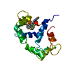
Entry Database : PDB / ID : 7pszTitle Crystal structure of CaM in complex with CDZ (form 1) Calmodulin-1 Keywords / / Function / homology Function Domain/homology Component
/ / / / / / / / / / / / / / / / / / / / / / / / / / / / / / / / / / / / / / / / / / / / / / / / / / / / / / / / / / / / / / / / / / / / / / / / / / / / / / / / / / / / / / / / / / / / / / / / / / / / / / / / / / / / / / / / / / / / / / / / / / / / / / / / Biological species Homo sapiens (human)Method / / / Resolution : 1.898 Å Authors Mechaly, A.E. / Leger, C. / Haouz, A. / Chenal, A. Funding support Organization Grant number Country Agence Nationale de la Recherche (ANR) Centre National de la Recherche Scientifique (CNRS)
Journal : Bmc Biol. / Year : 2022Title : Dynamics and structural changes of calmodulin upon interaction with the antagonist calmidazolium.Authors : Leger, C. / Pitard, I. / Sadi, M. / Carvalho, N. / Brier, S. / Mechaly, A. / Raoux-Barbot, D. / Davi, M. / Hoos, S. / Weber, P. / Vachette, P. / Durand, D. / Haouz, A. / Guijarro, J.I. / Ladant, D. / Chenal, A. History Deposition Sep 24, 2021 Deposition site / Processing site Revision 1.0 Aug 17, 2022 Provider / Type Revision 1.1 Jan 31, 2024 Group / Refinement descriptionCategory / chem_comp_bond / pdbx_initial_refinement_model
Show all Show less
 Movie
Movie Controller


 Open data
 Basic information
 Components
 Keywords
 Function and homology information
 Homo sapiens (human)
 X-RAY DIFFRACTION /
  Authors
 France, 2items
  Citation
 Structure visualization
 Downloads & links
 Other downloads
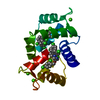
 PDBj
PDBj


 Assembly

 Mass: 652.245 Da / Num. of mol.: 1 / Source method: obtained synthetically / Formula: C31H23Cl6N2O / Feature type: SUBJECT OF INVESTIGATION
 Mass: 96.063 Da / Num. of mol.: 1 / Source method: obtained synthetically / Formula: SO4
 Mass: 40.078 Da / Num. of mol.: 4 / Source method: obtained synthetically / Formula: Ca
 Mass: 18.015 Da / Num. of mol.: 41 / Source method: isolated from a natural source / Formula: H2O
 Sample preparation
 Processing