 Movie
Movie Controller

[English] 日本語
 Yorodumi
- PDB-7prr: Structure of the ligand binding domain of the PctD (PA4633) chemo... -
+ Open data

- Basic information
| Entry | Database: PDB / ID: 7prr | |||||||||
|---|---|---|---|---|---|---|---|---|---|---|
| Title | Structure of the ligand binding domain of the PctD (PA4633) chemoreceptor of Pseudomonas aeruginosa PAO1 in complex with acetylcholine | |||||||||
 Components Components | Probable chemotaxis transducer | |||||||||
 Keywords Keywords | SIGNALING PROTEIN / Ligand binding domain / Pseudomonas aeruginosa / chemotactic transducer | |||||||||
| Function / homology |  Function and homology information Function and homology information | |||||||||
| Biological species |   Pseudomonas aeruginosa (bacteria) Pseudomonas aeruginosa (bacteria) | |||||||||
| Method |  X-RAY DIFFRACTION / SYNCHROTRON / MOLECULAR REPLACEMENT / Resolution: 1.8 Å X-RAY DIFFRACTION / SYNCHROTRON / MOLECULAR REPLACEMENT / Resolution: 1.8 Å | |||||||||
 Authors Authors | Gavira, J.A. / Matilla, M.A. / Martin-Mora, D. / Krell, T. | |||||||||
| Funding support |  Spain, 2items Spain, 2items
| |||||||||
 Citation Citation | Journal: Mbio / Year: 2022 Title: Chemotaxis of the Human Pathogen Pseudomonas aeruginosa to the Neurotransmitter Acetylcholine. Authors: Matilla, M.A. / Velando, F. / Tajuelo, A. / Martin-Mora, D. / Xu, W. / Sourjik, V. / Gavira, J.A. / Krell, T. | |||||||||
| History |
|
- Structure visualization
| Structure viewer | Molecule: MolmilJmol/JSmol |
|---|
- Downloads & links
-Download
| PDBx/mmCIF format | 7prr.cif.gz | 395.6 KB | Display | PDBx/mmCIF format |
|---|---|---|---|---|
| PDB format | pdb7prr.ent.gz | 315.5 KB | Display | PDB format |
| PDBx/mmJSON format | 7prr.json.gz | Tree view | PDBx/mmJSON format | |
| Others |  Other downloads Other downloads |
-Validation report
| Arichive directory | https://data.pdbj.org/pub/pdb/validation_reports/pr/7prrftp://data.pdbj.org/pub/pdb/validation_reports/pr/7prr | HTTPS FTP |
|---|
-Related structure data
| Related structure data |  7prqSC  7psgC S: Starting model for refinement C: citing same article ( |
|---|---|
| Similar structure data |
-Links
 PDBj
PDBj

- Assembly
| Deposited unit | 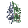
| ||||||||||||
|---|---|---|---|---|---|---|---|---|---|---|---|---|---|
| 1 |
| ||||||||||||
| Unit cell |
|
-Components
-Protein , 1 types, 2 molecules AB
| #1: Protein | Mass: 38453.246 Da / Num. of mol.: 2 Source method: isolated from a genetically manipulated source Details: Ligand binding domain Source: (gene. exp.) Pseudomonas aeruginosa (strain ATCC 15692 / DSM 22644 / CIP 104116 / JCM 14847 / LMG 12228 / 1C / PRS 101 / PAO1) (bacteria)Strain: ATCC 15692 / DSM 22644 / CIP 104116 / JCM 14847 / LMG 12228 / 1C / PRS 101 / PAO1 Gene: PA4633 / Plasmid: PET28B+ Production host: References: UniProt: Q9HVF8 |
|---|
-Non-polymers , 5 types, 460 molecules 
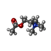


| #2: Chemical | ChemComp-PGE /  Mass: 150.173 Da / Num. of mol.: 4 / Source method: obtained synthetically / Formula: C6H14O4 Mass: 150.173 Da / Num. of mol.: 4 / Source method: obtained synthetically / Formula: C6H14O4#3: Chemical |  Mass: 146.207 Da / Num. of mol.: 2 / Source method: obtained synthetically / Formula: C7H16NO2 / Feature type: SUBJECT OF INVESTIGATION / Comment: neurotransmitter*YM Mass: 146.207 Da / Num. of mol.: 2 / Source method: obtained synthetically / Formula: C7H16NO2 / Feature type: SUBJECT OF INVESTIGATION / Comment: neurotransmitter*YM#4: Chemical | ChemComp-CA / |  Mass: 40.078 Da / Num. of mol.: 1 / Source method: obtained synthetically / Formula: Ca Mass: 40.078 Da / Num. of mol.: 1 / Source method: obtained synthetically / Formula: Ca#5: Chemical | ChemComp-PEG / |  Mass: 106.120 Da / Num. of mol.: 1 / Source method: obtained synthetically / Formula: C4H10O3 Mass: 106.120 Da / Num. of mol.: 1 / Source method: obtained synthetically / Formula: C4H10O3#6: Water | ChemComp-HOH / | Mass: 18.015 Da / Num. of mol.: 452 / Source method: isolated from a natural source / Formula: H2O |
|---|
-Details
| Has ligand of interest | Y |
|---|---|
| Has protein modification | Y |
-Experimental details
-Experiment
| Experiment | Method: X-RAY DIFFRACTION / Number of used crystals: 1 |
|---|
- Sample preparation
| Crystal | Density Matthews: 2.13 Å3/Da / Density % sol: 42.24 % |
|---|---|
| Crystal grow | Temperature: 293 K / Method: counter-diffusion / pH: 4.6 Details: 0.2 M Ammonium acetate, 0.1 M Sodium acetate trihydrate pH 4.6, 30% w/v Polyethylene glycol 4,000 |
-Data collection
| Diffraction | Mean temperature: 100 K / Serial crystal experiment: N | ||||||||||||||||||||||||||||||
|---|---|---|---|---|---|---|---|---|---|---|---|---|---|---|---|---|---|---|---|---|---|---|---|---|---|---|---|---|---|---|---|
| Diffraction source | Source: SYNCHROTRON / Site: ESRF  / Beamline: ID23-1 / Wavelength: 0.9763 Å / Beamline: ID23-1 / Wavelength: 0.9763 Å | ||||||||||||||||||||||||||||||
| Detector | Type: DECTRIS PILATUS 6M / Detector: PIXEL / Date: Feb 24, 2021 | ||||||||||||||||||||||||||||||
| Radiation | Protocol: SINGLE WAVELENGTH / Monochromatic (M) / Laue (L): M / Scattering type: x-ray | ||||||||||||||||||||||||||||||
| Radiation wavelength | Wavelength: 0.9763 Å / Relative weight: 1 | ||||||||||||||||||||||||||||||
| Reflection | Resolution: 1.8→47.48 Å / Num. obs: 62249 / % possible obs: 99 % / Redundancy: 4.5 % / CC1/2: 0.998 / Rmerge(I) obs: 0.086 / Rpim(I) all: 0.043 / Rrim(I) all: 0.096 / Net I/σ(I): 10.7 / Num. measured all: 280823 / Scaling rejects: 11 | ||||||||||||||||||||||||||||||
| Reflection shell | Diffraction-ID: 1
|
- Processing
| Software |
| ||||||||||||||||||||||||||||||||||||||||||||||||||||||||||||||||||||||||||||||||||||||||||||||||||||||||||||||||||||||||||||||||||||||||||||||||||||||||||||||||||||||||
|---|---|---|---|---|---|---|---|---|---|---|---|---|---|---|---|---|---|---|---|---|---|---|---|---|---|---|---|---|---|---|---|---|---|---|---|---|---|---|---|---|---|---|---|---|---|---|---|---|---|---|---|---|---|---|---|---|---|---|---|---|---|---|---|---|---|---|---|---|---|---|---|---|---|---|---|---|---|---|---|---|---|---|---|---|---|---|---|---|---|---|---|---|---|---|---|---|---|---|---|---|---|---|---|---|---|---|---|---|---|---|---|---|---|---|---|---|---|---|---|---|---|---|---|---|---|---|---|---|---|---|---|---|---|---|---|---|---|---|---|---|---|---|---|---|---|---|---|---|---|---|---|---|---|---|---|---|---|---|---|---|---|---|---|---|---|---|---|---|---|
| Refinement | Method to determine structure: MOLECULAR REPLACEMENT Starting model: 7PRQ Resolution: 1.8→47.48 Å / SU ML: 0.2018 / Cross valid method: FREE R-VALUE / σ(F): 1.34 / Phase error: 21.8005 Stereochemistry target values: GeoStd + Monomer Library + CDL v1.2
| ||||||||||||||||||||||||||||||||||||||||||||||||||||||||||||||||||||||||||||||||||||||||||||||||||||||||||||||||||||||||||||||||||||||||||||||||||||||||||||||||||||||||
| Solvent computation | Shrinkage radii: 0.9 Å / VDW probe radii: 1.11 Å / Solvent model: FLAT BULK SOLVENT MODEL | ||||||||||||||||||||||||||||||||||||||||||||||||||||||||||||||||||||||||||||||||||||||||||||||||||||||||||||||||||||||||||||||||||||||||||||||||||||||||||||||||||||||||
| Displacement parameters | Biso mean: 36.09 Å2 | ||||||||||||||||||||||||||||||||||||||||||||||||||||||||||||||||||||||||||||||||||||||||||||||||||||||||||||||||||||||||||||||||||||||||||||||||||||||||||||||||||||||||
| Refinement step | Cycle: LAST / Resolution: 1.8→47.48 Å
| ||||||||||||||||||||||||||||||||||||||||||||||||||||||||||||||||||||||||||||||||||||||||||||||||||||||||||||||||||||||||||||||||||||||||||||||||||||||||||||||||||||||||
| Refine LS restraints |
| ||||||||||||||||||||||||||||||||||||||||||||||||||||||||||||||||||||||||||||||||||||||||||||||||||||||||||||||||||||||||||||||||||||||||||||||||||||||||||||||||||||||||
| LS refinement shell |
|
