| Entry | Database: PDB / ID: 7ppt
|
|---|
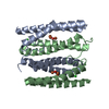
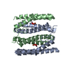
| Title | Structure of diFe-Sulerythrin at 0.26 MGy total absorbed dose |
|---|
 Components Components | Sulerythrin |
|---|
 Keywords Keywords | ELECTRON TRANSPORT / radiation damage / spatially resolved anomalous dispersion refinement / redox reaction |
|---|
| Function / homology |  Function and homology information Function and homology information
: / Rubrerythrin, diiron-binding domain / Rubrerythrin / Ferritin-like diiron domain / Ferritin-like diiron domain profile. / Ferritin-like / Ferritin-like superfamilySimilarity search - Domain/homology |
|---|
| Biological species |   Sulfurisphaera tokodaii str. 7 (archaea) Sulfurisphaera tokodaii str. 7 (archaea) |
|---|
| Method |  X-RAY DIFFRACTION / SYNCHROTRON / MOLECULAR REPLACEMENT / molecular replacement / Resolution: 1.42 Å X-RAY DIFFRACTION / SYNCHROTRON / MOLECULAR REPLACEMENT / molecular replacement / Resolution: 1.42 Å |
|---|
 Authors Authors | Lennartz, F. / Weiss, M.S. |
|---|
| Funding support |  Germany, 1items Germany, 1items | Organization | Grant number | Country |
|---|
| German Research Foundation (DFG) | 390540038 | Germany |
|
|---|
 Citation Citation | Journal: Acta Crystallogr D Struct Biol / Year: 2022
Title: Determining the oxidation state of elements by X-ray crystallography.
Authors: Lennartz, F. / Jeoung, J.H. / Ruenger, S. / Dobbek, H. / Weiss, M.S. |
|---|
| History | | Deposition | Sep 15, 2021 | Deposition site: PDBE / Processing site: PDBE |
|---|
| Revision 1.0 | Jun 8, 2022 | Provider: repository / Type: Initial release |
|---|
| Revision 1.1 | Jan 31, 2024 | Group: Data collection / Refinement description
Category: chem_comp_atom / chem_comp_bond / pdbx_initial_refinement_model |
|---|
|
|---|
 Movie
Movie Controller


 Open data
 Basic information
 Structure visualization
 Downloads & links
 Other downloads
 PDBj
PDBj


 Assembly