 Movie
Movie Controller

[English] 日本語
 Yorodumi
- PDB-7pom: Three dimensional structure of human carbonic anhydrase IX in com... -
+ Open data

- Basic information
| Entry | Database: PDB / ID: 7pom | ||||||
|---|---|---|---|---|---|---|---|
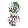
| Title | Three dimensional structure of human carbonic anhydrase IX in complex with sulfonamide | ||||||
 Components Components | Carbonic anhydrase 9 | ||||||
 Keywords Keywords | LYASE / CA IX / CA 9 / carbonic anhydrase IX / carbonic anhydrase 9 | ||||||
| Function / homology |  Function and homology information Function and homology informationRegulation of gene expression by Hypoxia-inducible Factor / response to testosterone / microvillus membrane / secretion / molecular function activator activity / Reversible hydration of carbon dioxide / morphogenesis of an epithelium / carbonic anhydrase / carbonate dehydratase activity / basolateral plasma membrane ...Regulation of gene expression by Hypoxia-inducible Factor / response to testosterone / microvillus membrane / secretion / molecular function activator activity / Reversible hydration of carbon dioxide / morphogenesis of an epithelium / carbonic anhydrase / carbonate dehydratase activity / basolateral plasma membrane / response to hypoxia / response to xenobiotic stimulus / nucleolus / zinc ion binding / membrane / plasma membrane Similarity search - Function | ||||||
| Biological species |  Homo sapiens (human) Homo sapiens (human) | ||||||
| Method |  X-RAY DIFFRACTION / SYNCHROTRON / MOLECULAR REPLACEMENT / Resolution: 1.98 Å X-RAY DIFFRACTION / SYNCHROTRON / MOLECULAR REPLACEMENT / Resolution: 1.98 Å | ||||||
 Authors Authors | Leitans, J. / Tars, K. | ||||||
| Funding support | 1items
| ||||||
 Citation Citation | Journal: Int J Mol Sci / Year: 2021 Title: Methyl 2-Halo-4-Substituted-5-Sulfamoyl-Benzoates as High Affinity and Selective Inhibitors of Carbonic Anhydrase IX. Authors: Zaksauskas, A. / Capkauskaite, E. / Paketuryte-Latve, V. / Smirnov, A. / Leitans, J. / Kazaks, A. / Dvinskis, E. / Stancaitis, L. / Mickeviciute, A. / Jachno, J. / Jezepcikas, L. / ...Authors: Zaksauskas, A. / Capkauskaite, E. / Paketuryte-Latve, V. / Smirnov, A. / Leitans, J. / Kazaks, A. / Dvinskis, E. / Stancaitis, L. / Mickeviciute, A. / Jachno, J. / Jezepcikas, L. / Linkuviene, V. / Sakalauskas, A. / Manakova, E. / Grazulis, S. / Matuliene, J. / Tars, K. / Matulis, D. | ||||||
| History |
|
- Structure visualization
| Structure viewer | Molecule: MolmilJmol/JSmol |
|---|
- Downloads & links
-Download
| PDBx/mmCIF format | 7pom.cif.gz | 222.6 KB | Display | PDBx/mmCIF format |
|---|---|---|---|---|
| PDB format | pdb7pom.ent.gz | 176.5 KB | Display | PDB format |
| PDBx/mmJSON format | 7pom.json.gz | Tree view | PDBx/mmJSON format | |
| Others |  Other downloads Other downloads |
-Validation report
| Arichive directory | https://data.pdbj.org/pub/pdb/validation_reports/po/7pomftp://data.pdbj.org/pub/pdb/validation_reports/po/7pom | HTTPS FTP |
|---|
-Related structure data
| Related structure data | 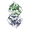 7pp9C 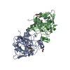 7puuC 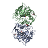 7puvC  7puwC 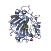 7q0cC  7q0dC 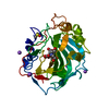 7q0eC  6fe2S S: Starting model for refinement C: citing same article ( |
|---|---|
| Similar structure data |


-Links
 PDBj
PDBj


- Assembly
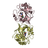
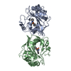
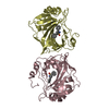
| Deposited unit | 
| ||||||||
|---|---|---|---|---|---|---|---|---|---|
| 1 | 
| ||||||||
| 2 | 
| ||||||||
| Unit cell |
|
-Components
| #1: Protein | Mass: 28186.711 Da / Num. of mol.: 4 / Mutation: C41S, N213Q Source method: isolated from a genetically manipulated source Details: Two dimers / Source: (gene. exp.) Homo sapiens (human) / Gene: CA9, G250, MN / Cell line (production host): Pichia pastoris / Production host:  Komagataella pastoris (fungus) / References: UniProt: Q16790, carbonic anhydrase Komagataella pastoris (fungus) / References: UniProt: Q16790, carbonic anhydrase#2: Chemical | ChemComp-ZN /   Mass: 65.409 Da / Num. of mol.: 4 / Source method: obtained synthetically / Formula: Zn Mass: 65.409 Da / Num. of mol.: 4 / Source method: obtained synthetically / Formula: Zn#3: Chemical | ChemComp-7VZ /   Mass: 363.880 Da / Num. of mol.: 4 / Source method: obtained synthetically / Formula: C14H18ClNO4S2 / Feature type: SUBJECT OF INVESTIGATION Mass: 363.880 Da / Num. of mol.: 4 / Source method: obtained synthetically / Formula: C14H18ClNO4S2 / Feature type: SUBJECT OF INVESTIGATION#4: Water | ChemComp-HOH / |  Mass: 18.015 Da / Num. of mol.: 741 / Source method: isolated from a natural source / Formula: H2O Mass: 18.015 Da / Num. of mol.: 741 / Source method: isolated from a natural source / Formula: H2OHas ligand of interest | Y | Has protein modification | Y | |
|---|
-Experimental details
-Experiment
| Experiment | Method: X-RAY DIFFRACTION / Number of used crystals: 1 |
|---|
- Sample preparation
| Crystal | Density Matthews: 3.44 Å3/Da / Density % sol: 64.22 % |
|---|---|
| Crystal grow | Temperature: 294 K / Method: vapor diffusion, sitting drop / pH: 4.5 Details: CRYSTALLIZATION CONDITIONS: 1.0 M DI-AMMONIUM HYDROGEN PHOSPHATE, 0.1 M SODIUM ACETATE PH 4.5, PROTEIN 10 MG/ML, 5-10 MM INHIBITOR (STOCK SOLUTION WAS 100 MM INHIBITOR DISSOLVED IN 100% ...Details: CRYSTALLIZATION CONDITIONS: 1.0 M DI-AMMONIUM HYDROGEN PHOSPHATE, 0.1 M SODIUM ACETATE PH 4.5, PROTEIN 10 MG/ML, 5-10 MM INHIBITOR (STOCK SOLUTION WAS 100 MM INHIBITOR DISSOLVED IN 100% DIMETHYL SULFOXIDE), VAPOR DIFFUSION, SITTING DROP, TEMPERATURE 294K |
-Data collection
| Diffraction | Mean temperature: 100 K / Serial crystal experiment: N |
|---|---|
| Diffraction source | Source: SYNCHROTRON / Site: BESSY  / Beamline: 14.1 / Wavelength: 0.9184 Å / Beamline: 14.1 / Wavelength: 0.9184 Å |
| Detector | Type: DECTRIS PILATUS 6M / Detector: PIXEL / Date: Jun 27, 2016 |
| Radiation | Monochromator: KMC-1 / Protocol: SINGLE WAVELENGTH / Monochromatic (M) / Laue (L): M / Scattering type: x-ray |
| Radiation wavelength | Wavelength: 0.9184 Å / Relative weight: 1 |
| Reflection | Resolution: 1.98→36.01 Å / Num. obs: 101441 / % possible obs: 97 % / Redundancy: 2.7 % / Rmerge(I) obs: 0.081 / Net I/σ(I): 6.7 |
| Reflection shell | Resolution: 1.98→2.08 Å / Redundancy: 2.8 % / Rmerge(I) obs: 0.543 / Mean I/σ(I) obs: 2 / Num. unique obs: 15100 / % possible all: 98.7 |
- Processing
| Software |
| ||||||||||||||||||||||||||||||||||||||||||||||||||||||||||||
|---|---|---|---|---|---|---|---|---|---|---|---|---|---|---|---|---|---|---|---|---|---|---|---|---|---|---|---|---|---|---|---|---|---|---|---|---|---|---|---|---|---|---|---|---|---|---|---|---|---|---|---|---|---|---|---|---|---|---|---|---|---|
| Refinement | Method to determine structure: MOLECULAR REPLACEMENT Starting model: 6FE2 Resolution: 1.98→36.01 Å / Cor.coef. Fo:Fc: 0.961 / Cor.coef. Fo:Fc free: 0.947 / SU B: 3.91 / SU ML: 0.104 / Cross valid method: THROUGHOUT / σ(F): 0 / ESU R: 0.132 / ESU R Free: 0.128 / Stereochemistry target values: MAXIMUM LIKELIHOOD Details: HYDROGENS HAVE BEEN ADDED IN THE RIDING POSITIONS U VALUES, REFINED INDIVIDUALLY
| ||||||||||||||||||||||||||||||||||||||||||||||||||||||||||||
| Solvent computation | Ion probe radii: 0.8 Å / Shrinkage radii: 0.8 Å / VDW probe radii: 1.2 Å / Solvent model: MASK | ||||||||||||||||||||||||||||||||||||||||||||||||||||||||||||
| Displacement parameters | Biso max: 137.19 Å2 / Biso mean: 33.736 Å2 / Biso min: 16.01 Å2
| ||||||||||||||||||||||||||||||||||||||||||||||||||||||||||||
| Refinement step | Cycle: final / Resolution: 1.98→36.01 Å
| ||||||||||||||||||||||||||||||||||||||||||||||||||||||||||||
| Refine LS restraints |
| ||||||||||||||||||||||||||||||||||||||||||||||||||||||||||||
| LS refinement shell | Resolution: 1.98→2.026 Å / Rfactor Rfree error: 0
|
