 Movie
Movie Controller

[English] 日本語
 Yorodumi
- PDB-7pjc: The structure of Candida albicans phosphoglucomutase with isothia... -
+ Open data

- Basic information
| Entry | Database: PDB / ID: 7pjc | |||||||||
|---|---|---|---|---|---|---|---|---|---|---|
| Title | The structure of Candida albicans phosphoglucomutase with isothiazolone modification on Cys359 | |||||||||
 Components Components | Phosphoglucomutase | |||||||||
 Keywords Keywords | ISOMERASE / phosphoglucomutase / candida / fungi | |||||||||
| Function / homology | Alpha-D-Glucose-1,6-Bisphosphate; Chain A, domain 3 / Alpha-D-Glucose-1,6-Bisphosphate, subunit A, domain 3 / Alpha-D-phosphohexomutase, C-terminal domain / TATA-Binding Protein / 2-Layer Sandwich / 3-Layer(aba) Sandwich / Alpha Beta / Chem-A4W Function and homology information Function and homology information | |||||||||
| Biological species |  Candida albicans SC5314 (yeast) Candida albicans SC5314 (yeast) | |||||||||
| Method |  X-RAY DIFFRACTION / SYNCHROTRON / MOLECULAR REPLACEMENT / Resolution: 2.11 Å X-RAY DIFFRACTION / SYNCHROTRON / MOLECULAR REPLACEMENT / Resolution: 2.11 Å | |||||||||
 Authors Authors | Yan, K. / van Aalten, D.M.F. | |||||||||
| Funding support |  United Kingdom, 2items United Kingdom, 2items
| |||||||||
 Citation Citation | Journal: To Be Published Title: Targeting an essential step in the biosynthetic pathway of uridine diphosphate glucose in Aspergillus fumigatus Authors: Yan, K. / Stanley, M. / Kowalski, B. / Raimi, O.G. / Ferenbach, A.T. / Wei, P. / Yuan, H. / Fang, W. / van Aalten, D.M.F. | |||||||||
| History |
|
- Structure visualization
| Structure viewer | Molecule: MolmilJmol/JSmol |
|---|
- Downloads & links
-Download
| PDBx/mmCIF format | 7pjc.cif.gz | 461.1 KB | Display | PDBx/mmCIF format |
|---|---|---|---|---|
| PDB format | pdb7pjc.ent.gz | 374.8 KB | Display | PDB format |
| PDBx/mmJSON format | 7pjc.json.gz | Tree view | PDBx/mmJSON format | |
| Others |  Other downloads Other downloads |
-Validation report
| Arichive directory | https://data.pdbj.org/pub/pdb/validation_reports/pj/7pjcftp://data.pdbj.org/pub/pdb/validation_reports/pj/7pjc | HTTPS FTP |
|---|
-Related structure data
| Related structure data | 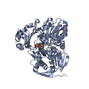 7p5oC 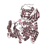 7pizSC S: Starting model for refinement C: citing same article ( |
|---|---|
| Similar structure data |
-Links
 PDBj
PDBj- Assembly
| Deposited unit | 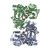
| ||||||||
|---|---|---|---|---|---|---|---|---|---|
| 1 | 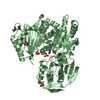
| ||||||||
| 2 | 
| ||||||||
| Unit cell |
|
-Components
| #1: Protein | Mass: 62290.051 Da / Num. of mol.: 2 Source method: isolated from a genetically manipulated source Details: Gly -4 Expression tag Pro -3 Expression tag Leu -2 Expression tag Gly -1 Restriction site Ser 0 Restriction site Source: (gene. exp.) Candida albicans SC5314 (yeast) / Strain: SC5314 / ATCC MYA-2876 / Gene: PGM2, CAALFM_CR02820WA, orf19.2841 / Production host:  References: phosphoglucomutase (alpha-D-glucose-1,6-bisphosphate-dependent) #2: Chemical |   Mass: 233.690 Da / Num. of mol.: 2 / Source method: obtained synthetically / Formula: C9H9ClFNOS Mass: 233.690 Da / Num. of mol.: 2 / Source method: obtained synthetically / Formula: C9H9ClFNOS#3: Chemical | ChemComp-SO4 /   Mass: 96.063 Da / Num. of mol.: 10 / Source method: obtained synthetically / Formula: SO4 Mass: 96.063 Da / Num. of mol.: 10 / Source method: obtained synthetically / Formula: SO4#4: Chemical | ChemComp-GOL / |   Mass: 92.094 Da / Num. of mol.: 1 / Source method: obtained synthetically / Formula: C3H8O3 Mass: 92.094 Da / Num. of mol.: 1 / Source method: obtained synthetically / Formula: C3H8O3#5: Water | ChemComp-HOH / |  Mass: 18.015 Da / Num. of mol.: 627 / Source method: isolated from a natural source / Formula: H2O Mass: 18.015 Da / Num. of mol.: 627 / Source method: isolated from a natural source / Formula: H2OHas ligand of interest | N | Has protein modification | Y | |
|---|
-Experimental details
-Experiment
| Experiment | Method: X-RAY DIFFRACTION / Number of used crystals: 1 |
|---|
- Sample preparation
| Crystal | Density Matthews: 2.56 Å3/Da / Density % sol: 52.04 % |
|---|---|
| Crystal grow | Temperature: 288.15 K / Method: vapor diffusion, sitting drop Details: 200 mM lithium sulphate monohydrate, 100 mM Tris-HCl pH 8.5, 30% w/v polyethylene glycol 4,000 |
-Data collection
| Diffraction | Mean temperature: 100 K / Serial crystal experiment: N |
|---|---|
| Diffraction source | Source: SYNCHROTRON / Site: Diamond / Beamline: I04 / Wavelength: 1.0332 Å |
| Detector | Type: DECTRIS EIGER2 XE 16M / Detector: PIXEL / Date: Nov 28, 2018 |
| Radiation | Protocol: SINGLE WAVELENGTH / Monochromatic (M) / Laue (L): M / Scattering type: x-ray |
| Radiation wavelength | Wavelength: 1.0332 Å / Relative weight: 1 |
| Reflection | Resolution: 2.11→67.975 Å / Num. obs: 47519 / % possible obs: 93 % / Redundancy: 3.2 % / Biso Wilson estimate: 25.18 Å2 / CC1/2: 0.993 / Net I/σ(I): 4 |
| Reflection shell | Resolution: 2.11→2.339 Å / Num. unique obs: 2376 / CC1/2: 0.855 |
- Processing
| Software |
| ||||||||||||||||||||||||||||||||||||||||||||||||||||||||||||||||||||||||||||||||||||||||||||||||||||||||||||||||||||||||||||||
|---|---|---|---|---|---|---|---|---|---|---|---|---|---|---|---|---|---|---|---|---|---|---|---|---|---|---|---|---|---|---|---|---|---|---|---|---|---|---|---|---|---|---|---|---|---|---|---|---|---|---|---|---|---|---|---|---|---|---|---|---|---|---|---|---|---|---|---|---|---|---|---|---|---|---|---|---|---|---|---|---|---|---|---|---|---|---|---|---|---|---|---|---|---|---|---|---|---|---|---|---|---|---|---|---|---|---|---|---|---|---|---|---|---|---|---|---|---|---|---|---|---|---|---|---|---|---|---|
| Refinement | Method to determine structure: MOLECULAR REPLACEMENT Starting model: 7PIZ Resolution: 2.11→67.97 Å / SU ML: 0.25 / Cross valid method: THROUGHOUT / σ(F): 1.33 / Phase error: 34.66 / Stereochemistry target values: ML
| ||||||||||||||||||||||||||||||||||||||||||||||||||||||||||||||||||||||||||||||||||||||||||||||||||||||||||||||||||||||||||||||
| Solvent computation | Shrinkage radii: 0.9 Å / VDW probe radii: 1.11 Å / Solvent model: FLAT BULK SOLVENT MODEL | ||||||||||||||||||||||||||||||||||||||||||||||||||||||||||||||||||||||||||||||||||||||||||||||||||||||||||||||||||||||||||||||
| Displacement parameters | Biso max: 110.32 Å2 / Biso mean: 31.5784 Å2 / Biso min: 9.05 Å2 | ||||||||||||||||||||||||||||||||||||||||||||||||||||||||||||||||||||||||||||||||||||||||||||||||||||||||||||||||||||||||||||||
| Refinement step | Cycle: final / Resolution: 2.11→67.97 Å
| ||||||||||||||||||||||||||||||||||||||||||||||||||||||||||||||||||||||||||||||||||||||||||||||||||||||||||||||||||||||||||||||
| LS refinement shell | Refine-ID: X-RAY DIFFRACTION / Rfactor Rfree error: 0 / Total num. of bins used: 17
| ||||||||||||||||||||||||||||||||||||||||||||||||||||||||||||||||||||||||||||||||||||||||||||||||||||||||||||||||||||||||||||||
| Refinement TLS params. | Method: refined / Origin x: -7.2887 Å / Origin y: -0.193 Å / Origin z: -16.9365 Å
| ||||||||||||||||||||||||||||||||||||||||||||||||||||||||||||||||||||||||||||||||||||||||||||||||||||||||||||||||||||||||||||||
| Refinement TLS group |
|
