[English] 日本語
 Yorodumi
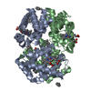
- PDB-7pcf: Human methemoglobin bound to Staphylococcus aureus hemophore IsdB -
+ Open data
- Basic information
| Entry | Database: PDB / ID: 7pcf | ||||||
|---|---|---|---|---|---|---|---|
| Title | Human methemoglobin bound to Staphylococcus aureus hemophore IsdB | ||||||
 Components Components |
| ||||||
 Keywords Keywords | METAL TRANSPORT / Iron acquisition / Hemophore / Hemoglobin / NEAT domain | ||||||
| Function / homology |  Function and homology information Function and homology informationheme transmembrane transporter activity / nitric oxide transport / hemoglobin alpha binding / cellular oxidant detoxification / hemoglobin binding / haptoglobin-hemoglobin complex / renal absorption / hemoglobin complex / oxygen transport / Scavenging of heme from plasma ...heme transmembrane transporter activity / nitric oxide transport / hemoglobin alpha binding / cellular oxidant detoxification / hemoglobin binding / haptoglobin-hemoglobin complex / renal absorption / hemoglobin complex / oxygen transport / Scavenging of heme from plasma / endocytic vesicle lumen / blood vessel diameter maintenance / oxygen carrier activity / hydrogen peroxide catabolic process / carbon dioxide transport / response to hydrogen peroxide / Heme signaling / Erythrocytes take up oxygen and release carbon dioxide / Erythrocytes take up carbon dioxide and release oxygen / Cytoprotection by HMOX1 / Late endosomal microautophagy / oxygen binding / regulation of blood pressure / platelet aggregation / Chaperone Mediated Autophagy / positive regulation of nitric oxide biosynthetic process / tertiary granule lumen / Factors involved in megakaryocyte development and platelet production / blood microparticle / ficolin-1-rich granule lumen / iron ion binding / inflammatory response / heme binding / Neutrophil degranulation / extracellular space / extracellular exosome / extracellular region / metal ion binding / membrane / cytosol Similarity search - Function | ||||||
| Biological species |  Staphylococcus aureus subsp. aureus MW2 (bacteria) Staphylococcus aureus subsp. aureus MW2 (bacteria) Homo sapiens (human) Homo sapiens (human) | ||||||
| Method | ELECTRON MICROSCOPY / single particle reconstruction / cryo EM / Resolution: 5.82 Å | ||||||
 Authors Authors | De Bei, O. / Gianquinto, E. / Chirgadze, D.Y. / Hardwick, S.W. / Spyrakis, F. / Luisi, B.F. / Campanini, B. | ||||||
| Funding support |  United Kingdom, 1items United Kingdom, 1items
| ||||||
 Citation Citation |  Journal: Proc Natl Acad Sci U S A / Year: 2022 Journal: Proc Natl Acad Sci U S A / Year: 2022Title: Cryo-EM structures of staphylococcal IsdB bound to human hemoglobin reveal the process of heme extraction. Authors: Omar De Bei / Marialaura Marchetti / Luca Ronda / Eleonora Gianquinto / Loretta Lazzarato / Dimitri Y Chirgadze / Steven W Hardwick / Lee R Cooper / Francesca Spyrakis / Ben F Luisi / ...Authors: Omar De Bei / Marialaura Marchetti / Luca Ronda / Eleonora Gianquinto / Loretta Lazzarato / Dimitri Y Chirgadze / Steven W Hardwick / Lee R Cooper / Francesca Spyrakis / Ben F Luisi / Barbara Campanini / Stefano Bettati /   Abstract: Iron surface determinant B (IsdB) is a hemoglobin (Hb) receptor essential for hemic iron acquisition by Staphylococcus aureus. Heme transfer to IsdB is possible from oxidized Hb (metHb), but ...Iron surface determinant B (IsdB) is a hemoglobin (Hb) receptor essential for hemic iron acquisition by Staphylococcus aureus. Heme transfer to IsdB is possible from oxidized Hb (metHb), but inefficient from Hb either bound to oxygen (oxyHb) or bound to carbon monoxide (HbCO), and encompasses a sequence of structural events that are currently poorly understood. By single-particle cryo-electron microscopy, we determined the structure of two IsdB:Hb complexes, representing key species along the heme extraction pathway. The IsdB:HbCO structure, at 2.9-Å resolution, provides a snapshot of the preextraction complex. In this early stage of IsdB:Hb interaction, the hemophore binds to the β-subunits of the Hb tetramer, exploiting a folding-upon-binding mechanism that is likely triggered by a cis/trans isomerization of Pro173. Binding of IsdB to α-subunits occurs upon dissociation of the Hb tetramer into α/β dimers. The structure of the IsdB:metHb complex reveals the final step of the extraction process, where heme transfer to IsdB is completed. The stability of the complex, both before and after heme transfer from Hb to IsdB, is influenced by isomerization of Pro173. These results greatly enhance current understanding of structural and dynamic aspects of the heme extraction mechanism by IsdB and provide insight into the interactions that stabilize the complex before the heme transfer event. This information will support future efforts to identify inhibitors of heme acquisition by S. aureus by interfering with IsdB:Hb complex formation. | ||||||
| History |
|
- Structure visualization
| Structure viewer | Molecule:  Molmil Molmil Jmol/JSmol Jmol/JSmol |
|---|
- Downloads & links
- Download
| PDBx/mmCIF format |  7pcf.cif.gz 7pcf.cif.gz | 151.5 KB | Display |  PDBx/mmCIF format PDBx/mmCIF format |
|---|---|---|---|---|
| PDB format |  pdb7pcf.ent.gz pdb7pcf.ent.gz | 99.8 KB | Display |  PDB format PDB format |
| PDBx/mmJSON format |  7pcf.json.gz 7pcf.json.gz | Tree view |  PDBx/mmJSON format PDBx/mmJSON format | |
| Others |  Other downloads Other downloads |
-Validation report
| Summary document |  7pcf_validation.pdf.gz 7pcf_validation.pdf.gz | 1.3 MB | Display |  wwPDB validaton report wwPDB validaton report |
|---|---|---|---|---|
| Full document |  7pcf_full_validation.pdf.gz 7pcf_full_validation.pdf.gz | 1.3 MB | Display | |
| Data in XML |  7pcf_validation.xml.gz 7pcf_validation.xml.gz | 34.6 KB | Display | |
| Data in CIF |  7pcf_validation.cif.gz 7pcf_validation.cif.gz | 55.7 KB | Display | |
| Arichive directory |  https://data.pdbj.org/pub/pdb/validation_reports/pc/7pcf https://data.pdbj.org/pub/pdb/validation_reports/pc/7pcf ftp://data.pdbj.org/pub/pdb/validation_reports/pc/7pcf ftp://data.pdbj.org/pub/pdb/validation_reports/pc/7pcf | HTTPS FTP |
-Related structure data
| Related structure data |  13319MC  7pchC  7pcqC M: map data used to model this data C: citing same article ( |
|---|---|
| Similar structure data | Similarity search - Function & homology  F&H Search F&H Search |
- Links
Links
- Assembly
| Deposited unit | 
|
|---|---|
| 1 |
|
- Components
| #1: Protein | Mass: 15150.353 Da / Num. of mol.: 1 / Source method: isolated from a natural source / Source: (natural)  Homo sapiens (human) / References: UniProt: P69905 Homo sapiens (human) / References: UniProt: P69905 | ||||
|---|---|---|---|---|---|
| #2: Protein | Mass: 15890.198 Da / Num. of mol.: 1 / Source method: isolated from a natural source / Source: (natural)  Homo sapiens (human) / References: UniProt: P68871 Homo sapiens (human) / References: UniProt: P68871 | ||||
| #3: Protein | Mass: 43393.129 Da / Num. of mol.: 2 Source method: isolated from a genetically manipulated source Source: (gene. exp.)  Staphylococcus aureus subsp. aureus MW2 (bacteria) Staphylococcus aureus subsp. aureus MW2 (bacteria)Gene: isdB, frpB, sasJ, sirH, MW1011 / Plasmid: pASK-IBA3plus / Production host:  #4: Chemical | Has ligand of interest | Y | |
-Experimental details
-Experiment
| Experiment | Method: ELECTRON MICROSCOPY |
|---|---|
| EM experiment | Aggregation state: PARTICLE / 3D reconstruction method: single particle reconstruction |
- Sample preparation
| Component |
| ||||||||||||||||||||||||
|---|---|---|---|---|---|---|---|---|---|---|---|---|---|---|---|---|---|---|---|---|---|---|---|---|---|
| Molecular weight | Value: 0.119 MDa / Experimental value: NO | ||||||||||||||||||||||||
| Source (natural) |
| ||||||||||||||||||||||||
| Source (recombinant) | Organism:  | ||||||||||||||||||||||||
| Buffer solution | pH: 7.2 Details: CHAPSO was added immediately before plunge freezing to overcome preferred orientation of the particles in the vitreous ice. | ||||||||||||||||||||||||
| Buffer component |
| ||||||||||||||||||||||||
| Specimen | Conc.: 2 mg/ml / Embedding applied: NO / Shadowing applied: NO / Staining applied: NO / Vitrification applied: YES | ||||||||||||||||||||||||
| Specimen support | Details: The grid was discharged on both sides. / Grid material: GOLD / Grid mesh size: 300 divisions/in. / Grid type: UltrAuFoil R1.2/1.3 | ||||||||||||||||||||||||
| Vitrification | Instrument: FEI VITROBOT MARK IV / Cryogen name: ETHANE / Humidity: 99 % / Chamber temperature: 277.15 K |
- Electron microscopy imaging
| Experimental equipment |  Model: Titan Krios / Image courtesy: FEI Company |
|---|---|
| Microscopy | Model: FEI TITAN KRIOS |
| Electron gun | Electron source:  FIELD EMISSION GUN / Accelerating voltage: 300 kV / Illumination mode: FLOOD BEAM FIELD EMISSION GUN / Accelerating voltage: 300 kV / Illumination mode: FLOOD BEAM |
| Electron lens | Mode: BRIGHT FIELD / Nominal magnification: 130000 X / Nominal defocus max: 3100 nm / Nominal defocus min: 1000 nm / Cs: 2.7 mm / C2 aperture diameter: 50 µm |
| Specimen holder | Cryogen: NITROGEN |
| Image recording | Electron dose: 39.59 e/Å2 / Film or detector model: GATAN K3 BIOQUANTUM (6k x 4k) / Num. of grids imaged: 1 / Num. of real images: 3051 |
| EM imaging optics | Energyfilter slit width: 20 eV |
- Processing
| EM software |
| |||||||||||||||||||||||||||||||||||||||||||||||||||||||
|---|---|---|---|---|---|---|---|---|---|---|---|---|---|---|---|---|---|---|---|---|---|---|---|---|---|---|---|---|---|---|---|---|---|---|---|---|---|---|---|---|---|---|---|---|---|---|---|---|---|---|---|---|---|---|---|---|
| CTF correction | Type: PHASE FLIPPING AND AMPLITUDE CORRECTION | |||||||||||||||||||||||||||||||||||||||||||||||||||||||
| Particle selection | Num. of particles selected: 521822 | |||||||||||||||||||||||||||||||||||||||||||||||||||||||
| Symmetry | Point symmetry: C1 (asymmetric) | |||||||||||||||||||||||||||||||||||||||||||||||||||||||
| 3D reconstruction | Resolution: 5.82 Å / Resolution method: FSC 0.143 CUT-OFF / Num. of particles: 103653 / Num. of class averages: 1 / Symmetry type: POINT | |||||||||||||||||||||||||||||||||||||||||||||||||||||||
| Atomic model building | Protocol: RIGID BODY FIT / Space: REAL | |||||||||||||||||||||||||||||||||||||||||||||||||||||||
| Atomic model building |
|
 Movie
Movie Controller


 PDBj
PDBj


