 Movie
Movie Controller

[English] 日本語
 Yorodumi
- PDB-7pca: Functional and structural characterization of redox sensitive sup... -
+ Open data

- Basic information
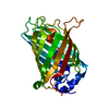
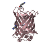
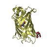
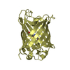
| Entry | Database: PDB / ID: 7pca | |||||||||
|---|---|---|---|---|---|---|---|---|---|---|
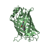
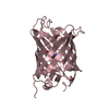
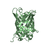
| Title | Functional and structural characterization of redox sensitive superfolder green fluorescent protein and variants | |||||||||
 Components Components | Green fluorescent protein | |||||||||
 Keywords Keywords | FLUORESCENT PROTEIN / genetically encoded biosensors / x-ray crystal structure / dynamic simulation / redox regulation / Plasmodium falciparum | |||||||||
| Function / homology | Green fluorescent protein, GFP / Green fluorescent protein-related / Green fluorescent protein / Green fluorescent protein / bioluminescence / generation of precursor metabolites and energy / FORMAMIDE / ETHANOL / Green fluorescent protein Function and homology information Function and homology information | |||||||||
| Biological species |   Aequorea victoria (jellyfish) Aequorea victoria (jellyfish) | |||||||||
| Method |  X-RAY DIFFRACTION / SYNCHROTRON / MOLECULAR REPLACEMENT / Resolution: 1.05 Å X-RAY DIFFRACTION / SYNCHROTRON / MOLECULAR REPLACEMENT / Resolution: 1.05 Å | |||||||||
 Authors Authors | Fritz-Wolf, K. / Heimsch, K.C. / Schuh, A.K. / Becker, K. | |||||||||
| Funding support |  Germany, 1items Germany, 1items
| |||||||||
 Citation Citation | Journal: Antioxid.Redox Signal. / Year: 2022 Title: Structure and Function of Redox-Sensitive Superfolder Green Fluorescent Protein Variant. Authors: Heimsch, K.C. / Gertzen, C.G.W. / Schuh, A.K. / Nietzel, T. / Rahlfs, S. / Przyborski, J.M. / Gohlke, H. / Schwarzlander, M. / Becker, K. / Fritz-Wolf, K. | |||||||||
| History |
|
- Structure visualization
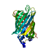
| Structure viewer | Molecule: MolmilJmol/JSmol |
|---|
- Downloads & links
-Download
| PDBx/mmCIF format | 7pca.cif.gz | 110.2 KB | Display | PDBx/mmCIF format |
|---|---|---|---|---|
| PDB format | pdb7pca.ent.gz | 83.2 KB | Display | PDB format |
| PDBx/mmJSON format | 7pca.json.gz | Tree view | PDBx/mmJSON format | |
| Others |  Other downloads Other downloads |
-Validation report
| Arichive directory | https://data.pdbj.org/pub/pdb/validation_reports/pc/7pcaftp://data.pdbj.org/pub/pdb/validation_reports/pc/7pca | HTTPS FTP |
|---|
-Related structure data
| Related structure data |  7pczC  7pd0C 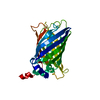 1qyoS S: Starting model for refinement C: citing same article ( |
|---|---|
| Similar structure data |


-Links
 PDBj
PDBj


- Assembly
| Deposited unit | 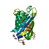
| ||||||||
|---|---|---|---|---|---|---|---|---|---|
| 1 |
| ||||||||
| Unit cell |
|
-Components
| #1: Protein | Mass: 26752.164 Da / Num. of mol.: 1 Source method: isolated from a genetically manipulated source Source: (gene. exp.) Aequorea victoria (jellyfish) / Gene: GFP / Production host:  | ||||||||||
|---|---|---|---|---|---|---|---|---|---|---|---|
| #2: Chemical |   Mass: 92.094 Da / Num. of mol.: 2 / Source method: obtained synthetically / Formula: C3H8O3 Mass: 92.094 Da / Num. of mol.: 2 / Source method: obtained synthetically / Formula: C3H8O3#3: Chemical |   Mass: 46.068 Da / Num. of mol.: 2 / Source method: obtained synthetically / Formula: C2H6O Mass: 46.068 Da / Num. of mol.: 2 / Source method: obtained synthetically / Formula: C2H6O#4: Chemical |   Type: L-peptide NH3 amino terminus / Mass: 45.041 Da / Num. of mol.: 2 / Source method: obtained synthetically / Formula: CH3NO Type: L-peptide NH3 amino terminus / Mass: 45.041 Da / Num. of mol.: 2 / Source method: obtained synthetically / Formula: CH3NO#5: Water | ChemComp-HOH / |  Mass: 18.015 Da / Num. of mol.: 281 / Source method: isolated from a natural source / Formula: H2O Mass: 18.015 Da / Num. of mol.: 281 / Source method: isolated from a natural source / Formula: H2OHas ligand of interest | Y | Has protein modification | Y | |
-Experimental details
-Experiment
| Experiment | Method: X-RAY DIFFRACTION / Number of used crystals: 1 |
|---|
- Sample preparation
| Crystal | Density Matthews: 1.92 Å3/Da / Density % sol: 36 % |
|---|---|
| Crystal grow | Temperature: 295 K / Method: vapor diffusion / pH: 7.5 / Details: 46% ETOH, 0.25% v/v Dichloromethane |
-Data collection
| Diffraction | Mean temperature: 100 K / Serial crystal experiment: N |
|---|---|
| Diffraction source | Source: SYNCHROTRON / Site: SLS  / Beamline: X10SA / Wavelength: 1 Å / Beamline: X10SA / Wavelength: 1 Å |
| Detector | Type: DECTRIS PILATUS 6M / Detector: PIXEL / Date: May 6, 2018 |
| Radiation | Protocol: SINGLE WAVELENGTH / Monochromatic (M) / Laue (L): M / Scattering type: x-ray |
| Radiation wavelength | Wavelength: 1 Å / Relative weight: 1 |
| Reflection | Resolution: 1.05→19.85 Å / Num. obs: 89058 / % possible obs: 91.69 % / Redundancy: 4.4 % / Biso Wilson estimate: 8.85 Å2 / CC1/2: 0.999 / Rmerge(I) obs: 0.04413 / Rpim(I) all: 0.0227 / Net I/σ(I): 16.24 |
| Reflection shell | Resolution: 1.05→1.088 Å / Redundancy: 2.1 % / Num. unique obs: 4957 / CC1/2: 0.886 / % possible all: 51.7 |
- Processing
| Software |
| |||||||||||||||||||||||||||||||||||||||||||||||||||||||||||||||||||||||||||||||||||||||||||||||||||||||||||||||||||||||||||||||||||||||||||||||||||||||||||||||||||||||||||||||||||||||||||||||||||||||||||||||||||||||||
|---|---|---|---|---|---|---|---|---|---|---|---|---|---|---|---|---|---|---|---|---|---|---|---|---|---|---|---|---|---|---|---|---|---|---|---|---|---|---|---|---|---|---|---|---|---|---|---|---|---|---|---|---|---|---|---|---|---|---|---|---|---|---|---|---|---|---|---|---|---|---|---|---|---|---|---|---|---|---|---|---|---|---|---|---|---|---|---|---|---|---|---|---|---|---|---|---|---|---|---|---|---|---|---|---|---|---|---|---|---|---|---|---|---|---|---|---|---|---|---|---|---|---|---|---|---|---|---|---|---|---|---|---|---|---|---|---|---|---|---|---|---|---|---|---|---|---|---|---|---|---|---|---|---|---|---|---|---|---|---|---|---|---|---|---|---|---|---|---|---|---|---|---|---|---|---|---|---|---|---|---|---|---|---|---|---|---|---|---|---|---|---|---|---|---|---|---|---|---|---|---|---|---|---|---|---|---|---|---|---|---|---|---|---|---|---|---|---|---|
| Refinement | Method to determine structure: MOLECULAR REPLACEMENT Starting model: 1qyo Resolution: 1.05→19.85 Å / SU ML: 0.07 / Cross valid method: FREE R-VALUE / σ(F): 1.36 / Phase error: 15.47 / Stereochemistry target values: ML
| |||||||||||||||||||||||||||||||||||||||||||||||||||||||||||||||||||||||||||||||||||||||||||||||||||||||||||||||||||||||||||||||||||||||||||||||||||||||||||||||||||||||||||||||||||||||||||||||||||||||||||||||||||||||||
| Solvent computation | Shrinkage radii: 0.9 Å / VDW probe radii: 1.11 Å / Solvent model: FLAT BULK SOLVENT MODEL | |||||||||||||||||||||||||||||||||||||||||||||||||||||||||||||||||||||||||||||||||||||||||||||||||||||||||||||||||||||||||||||||||||||||||||||||||||||||||||||||||||||||||||||||||||||||||||||||||||||||||||||||||||||||||
| Refinement step | Cycle: LAST / Resolution: 1.05→19.85 Å
| |||||||||||||||||||||||||||||||||||||||||||||||||||||||||||||||||||||||||||||||||||||||||||||||||||||||||||||||||||||||||||||||||||||||||||||||||||||||||||||||||||||||||||||||||||||||||||||||||||||||||||||||||||||||||
| Refine LS restraints |
| |||||||||||||||||||||||||||||||||||||||||||||||||||||||||||||||||||||||||||||||||||||||||||||||||||||||||||||||||||||||||||||||||||||||||||||||||||||||||||||||||||||||||||||||||||||||||||||||||||||||||||||||||||||||||
| LS refinement shell |
|
