 Movie
Movie Controller


+ Open data

- Basic information
| Entry | Database: PDB / ID: 7p9o | ||||||
|---|---|---|---|---|---|---|---|
| Title | Structure of E.coli RlmJ in complex with a SAM analogue (CA) | ||||||
 Components Components | Ribosomal RNA large subunit methyltransferase J | ||||||
 Keywords Keywords | RNA BINDING PROTEIN / RNA MTases / methyltransferase / SAM analogue / m6A / RNA binding / SAM conjugate / RlmJ / conjugate analogue / RNA recognition / bisubstrate analogue | ||||||
| Function / homology |  Function and homology information Function and homology information23S rRNA (adenine2030-N6)-methyltransferase / 23S rRNA (adenine(2030)-N(6))-methyltransferase activity / rRNA (adenine-N6-)-methyltransferase activity / carbon utilization / rRNA base methylation / RNA binding / cytosol Similarity search - Function | ||||||
| Biological species |  | ||||||
| Method |  X-RAY DIFFRACTION / SYNCHROTRON / MOLECULAR REPLACEMENT / molecular replacement / Resolution: 2.095 Å X-RAY DIFFRACTION / SYNCHROTRON / MOLECULAR REPLACEMENT / molecular replacement / Resolution: 2.095 Å | ||||||
 Authors Authors | Meynier, V. / Catala, M. / Oerum, S. / Barraud, P. / Tisne, C. | ||||||
| Funding support |  France, 1items France, 1items
| ||||||
 Citation Citation | Journal: Nucleic Acids Res. / Year: 2022 Title: Synthesis of RNA-cofactor conjugates and structural exploration of RNA recognition by an m6A RNA methyltransferase. Authors: Meynier, V. / Iannazzo, L. / Catala, M. / Oerum, S. / Braud, E. / Atdjian, C. / Barraud, P. / Fonvielle, M. / Tisne, C. / Etheve-Quelquejeu, M. | ||||||
| History |
|
- Structure visualization
| Structure viewer | Molecule: MolmilJmol/JSmol |
|---|
- Downloads & links
-Download
| PDBx/mmCIF format | 7p9o.cif.gz | 116.6 KB | Display | PDBx/mmCIF format |
|---|---|---|---|---|
| PDB format | pdb7p9o.ent.gz | 91.3 KB | Display | PDB format |
| PDBx/mmJSON format | 7p9o.json.gz | Tree view | PDBx/mmJSON format | |
| Others |  Other downloads Other downloads |
-Validation report
| Arichive directory | https://data.pdbj.org/pub/pdb/validation_reports/p9/7p9oftp://data.pdbj.org/pub/pdb/validation_reports/p9/7p9o | HTTPS FTP |
|---|
-Related structure data
| Related structure data | 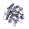 7p8qC 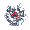 7p9iC 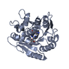 6qdxS S: Starting model for refinement C: citing same article ( |
|---|---|
| Similar structure data |
-Links
 PDBj
PDBj- Assembly
| Deposited unit | 
| ||||||||
|---|---|---|---|---|---|---|---|---|---|
| 1 |
| ||||||||
| Unit cell |
|
-Components
| #1: Protein | Mass: 31989.674 Da / Num. of mol.: 1 Source method: isolated from a genetically manipulated source Source: (gene. exp.) References: UniProt: P37634, 23S rRNA (adenine2030-N6)-methyltransferase |
|---|---|
| #2: Chemical | ChemComp-0Y0 /   Mass: 367.360 Da / Num. of mol.: 1 / Source method: obtained synthetically / Formula: C14H21N7O5 / Feature type: SUBJECT OF INVESTIGATION Mass: 367.360 Da / Num. of mol.: 1 / Source method: obtained synthetically / Formula: C14H21N7O5 / Feature type: SUBJECT OF INVESTIGATION |
| #3: Water | ChemComp-HOH /  Mass: 18.015 Da / Num. of mol.: 101 / Source method: isolated from a natural source / Formula: H2O Mass: 18.015 Da / Num. of mol.: 101 / Source method: isolated from a natural source / Formula: H2O |
| Has ligand of interest | Y |
-Experimental details
-Experiment
| Experiment | Method: X-RAY DIFFRACTION / Number of used crystals: 1 |
|---|
- Sample preparation
| Crystal | Density Matthews: 2.12 Å3/Da / Density % sol: 42.05 % |
|---|---|
| Crystal grow | Temperature: 293 K / Method: vapor diffusion / pH: 6.5 / Details: 0.1 M Bis-Tris pH 6.5 28% (w/v) PEG MME 2000 |
-Data collection
| Diffraction | Mean temperature: 100 K / Serial crystal experiment: N |
|---|---|
| Diffraction source | Source: SYNCHROTRON / Site: SOLEIL / Beamline: PROXIMA 1 / Wavelength: 0.9786 Å |
| Detector | Type: DECTRIS EIGER X 9M / Detector: PIXEL / Date: Dec 15, 2018 |
| Radiation | Monochromator: M / Protocol: SINGLE WAVELENGTH / Monochromatic (M) / Laue (L): M / Scattering type: x-ray |
| Radiation wavelength | Wavelength: 0.9786 Å / Relative weight: 1 |
| Reflection | Resolution: 2.095→43.651 Å / Num. obs: 15974 / % possible obs: 99.2 % / Redundancy: 6.7 % / CC1/2: 0.998 / Rmerge(I) obs: 0.083 / Rpim(I) all: 0.035 / Rrim(I) all: 0.091 / Net I/σ(I): 12.9 |
| Reflection shell | Resolution: 2.1→2.16 Å / Redundancy: 6.5 % / Rmerge(I) obs: 0.442 / Num. unique obs: 1224 / CC1/2: 0.966 / Rpim(I) all: 0.184 / Rrim(I) all: 0.479 / % possible all: 92.6 |
-Phasing
| Phasing | Method: molecular replacement |
|---|
- Processing
| Software |
| ||||||||||||||||||||||||||||||||||||
|---|---|---|---|---|---|---|---|---|---|---|---|---|---|---|---|---|---|---|---|---|---|---|---|---|---|---|---|---|---|---|---|---|---|---|---|---|---|
| Refinement | Method to determine structure: MOLECULAR REPLACEMENT Starting model: 6QDX Resolution: 2.095→43.651 Å / SU ML: 0.34 / Cross valid method: THROUGHOUT / σ(F): 1.37 / Phase error: 33.41 / Stereochemistry target values: ML
| ||||||||||||||||||||||||||||||||||||
| Solvent computation | Shrinkage radii: 0.9 Å / VDW probe radii: 1.11 Å / Solvent model: FLAT BULK SOLVENT MODEL | ||||||||||||||||||||||||||||||||||||
| Displacement parameters | Biso max: 68.86 Å2 / Biso mean: 22.7084 Å2 / Biso min: 2.43 Å2 | ||||||||||||||||||||||||||||||||||||
| Refinement step | Cycle: final / Resolution: 2.095→43.651 Å
| ||||||||||||||||||||||||||||||||||||
| LS refinement shell | Refine-ID: X-RAY DIFFRACTION / Rfactor Rfree error: 0
|
