regulation of starch biosynthetic process / regulation of chlorophyll biosynthetic process / oxidoreductase activity, acting on a sulfur group of donors, disulfide as acceptor / thioredoxin-disulfide reductase (NADPH) / thioredoxin-disulfide reductase (NADPH) activity / removal of superoxide radicals / cell redox homeostasis / chloroplast / hydrogen peroxide catabolic process / enzyme activator activity / nucleotide binding Similarity search - Function
Mass: 18.015 Da / Num. of mol.: 136 / Source method: isolated from a natural source / Formula: H2O
Has ligand of interest
Y
Has protein modification
Y
-
Experimental details
-
Experiment
Experiment
Method: X-RAY DIFFRACTION / Number of used crystals: 1
-
Sample preparation
Crystal grow
Temperature: 285.15 K / Method: vapor diffusion, hanging drop / pH: 8.5 Details: 0.1M mixture of DL-Glutamic acid monohydrate, DL-Alanine, Glycine, DL-Lysine monohydrochloride, DL-Serine; 0.1 M of Tris (base), BICINE with pH-8.5; 20% of v/v Ethylene glycol, 10 % of w/v PEG 8000
-
Data collection
Diffraction
Mean temperature: 100 K / Serial crystal experiment: N
Diffraction source
Source: SYNCHROTRON / Site: PETRA III, EMBL c/o DESY / Beamline: P13 (MX1) / Wavelength: 0.976 Å
In the structure databanks used in Yorodumi, some data are registered as the other names, "COVID-19 virus" and "2019-nCoV". Here are the details of the virus and the list of structure data.
Jan 31, 2019. EMDB accession codes are about to change! (news from PDBe EMDB page)
EMDB accession codes are about to change! (news from PDBe EMDB page)
The allocation of 4 digits for EMDB accession codes will soon come to an end. Whilst these codes will remain in use, new EMDB accession codes will include an additional digit and will expand incrementally as the available range of codes is exhausted. The current 4-digit format prefixed with “EMD-” (i.e. EMD-XXXX) will advance to a 5-digit format (i.e. EMD-XXXXX), and so on. It is currently estimated that the 4-digit codes will be depleted around Spring 2019, at which point the 5-digit format will come into force.
The EM Navigator/Yorodumi systems omit the EMD- prefix.
Related info.:Q: What is EMD? / ID/Accession-code notation in Yorodumi/EM Navigator
Yorodumi is a browser for structure data from EMDB, PDB, SASBDB, etc.
This page is also the successor to EM Navigator detail page, and also detail information page/front-end page for Omokage search.
The word "yorodu" (or yorozu) is an old Japanese word meaning "ten thousand". "mi" (miru) is to see.
Related info.:EMDB / PDB / SASBDB / Comparison of 3 databanks / Yorodumi Search / Aug 31, 2016. New EM Navigator & Yorodumi / Yorodumi Papers / Jmol/JSmol / Function and homology information / Changes in new EM Navigator and Yorodumi
 Movie
Movie Controller

 Yorodumi
 Open data
 Basic information
 Components
 Keywords
 Function and homology information
 Chlamydomonas reinhardtii (plant)
 X-RAY DIFFRACTION /
  Authors
 Germany, 1items
  Citation
 Structure visualization
 Downloads & links
 Other downloads


 PDBj
PDBj


 Assembly
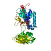
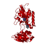


 Mass: 785.550 Da / Num. of mol.: 1 / Source method: obtained synthetically / Formula: C27H33N9O15P2 / Feature type: SUBJECT OF INVESTIGATION / Comment: FAD*YM
 Mass: 18.015 Da / Num. of mol.: 136 / Source method: isolated from a natural source / Formula: H2O
 Sample preparation
 Processing