[English] 日本語
Yorodumi
- PDB-7p8i: Receptor-binding domain (RBD) of the spike protein of the bat cor... -

+
Open data


ID or keywords:

Loading...

-
Basic information

Entry
Database: PDB / ID: 7p8i
Title
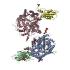
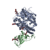
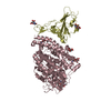
Receptor-binding domain (RBD) of the spike protein of the bat coronavirus RaTG13 virus in complex with the extracellular domain of human angiotensin-converting enzyme 2 (ACE2) - Crystal form 1
Components
  • Processed angiotensin-converting enzyme 2
  • Spike glycoproteinSpike protein
KeywordsCELL INVASION / COVID-19 Bat coronavirus Spillover Zoonotic viral infection Evolution / IMMUNE SYSTEM
Function / homology
Function and homology information


positive regulation of amino acid transport / angiotensin-converting enzyme 2 / positive regulation of L-proline import across plasma membrane / Hydrolases; Acting on peptide bonds (peptidases); Metallocarboxypeptidases / angiotensin-mediated drinking behavior / regulation of systemic arterial blood pressure by renin-angiotensin / tryptophan transport / positive regulation of gap junction assembly / regulation of vasoconstriction / regulation of cardiac conduction ...positive regulation of amino acid transport / angiotensin-converting enzyme 2 / positive regulation of L-proline import across plasma membrane / Hydrolases; Acting on peptide bonds (peptidases); Metallocarboxypeptidases / angiotensin-mediated drinking behavior / regulation of systemic arterial blood pressure by renin-angiotensin / tryptophan transport / positive regulation of gap junction assembly / regulation of vasoconstriction / regulation of cardiac conduction / peptidyl-dipeptidase activity / angiotensin maturation / maternal process involved in female pregnancy / metallocarboxypeptidase activity / Metabolism of Angiotensinogen to Angiotensins / Attachment and Entry / carboxypeptidase activity / negative regulation of signaling receptor activity / positive regulation of cardiac muscle contraction / regulation of cytokine production / viral life cycle / blood vessel diameter maintenance / brush border membrane / regulation of transmembrane transporter activity / negative regulation of smooth muscle cell proliferation / endocytosis involved in viral entry into host cell / cilium / negative regulation of ERK1 and ERK2 cascade / endocytic vesicle membrane / metallopeptidase activity / positive regulation of reactive oxygen species metabolic process / virus receptor activity / regulation of cell population proliferation / regulation of inflammatory response / endopeptidase activity / Induction of Cell-Cell Fusion / Potential therapeutics for SARS / host cell endoplasmic reticulum-Golgi intermediate compartment membrane / entry receptor-mediated virion attachment to host cell / receptor-mediated endocytosis of virus by host cell / Attachment and Entry / membrane fusion / receptor-mediated virion attachment to host cell / symbiont entry into host cell / membrane raft / apical plasma membrane / fusion of virus membrane with host plasma membrane / endoplasmic reticulum lumen / fusion of virus membrane with host endosome membrane / viral envelope / host cell plasma membrane / virion membrane / cell surface / extracellular space / extracellular exosome / zinc ion binding / extracellular region / membrane / identical protein binding / plasma membrane
Similarity search - Function
Collectrin-like domain profile. / Collectrin domain / Renal amino acid transporter / Peptidase family M2 domain profile. / Peptidase M2, peptidyl-dipeptidase A / Angiotensin-converting enzyme / Neutral zinc metallopeptidases, zinc-binding region signature. / Spike (S) protein S1 subunit, N-terminal domain, SARS-CoV-like / Betacoronavirus spike (S) glycoprotein S1 subunit N-terminal (NTD) domain profile. / Spike glycoprotein, N-terminal domain superfamily ...Collectrin-like domain profile. / Collectrin domain / Renal amino acid transporter / Peptidase family M2 domain profile. / Peptidase M2, peptidyl-dipeptidase A / Angiotensin-converting enzyme / Neutral zinc metallopeptidases, zinc-binding region signature. / Spike (S) protein S1 subunit, N-terminal domain, SARS-CoV-like / Betacoronavirus spike (S) glycoprotein S1 subunit N-terminal (NTD) domain profile. / Spike glycoprotein, N-terminal domain superfamily / Betacoronavirus spike (S) glycoprotein S1 subunit C-terminal (CTD) domain profile. / Spike glycoprotein, betacoronavirus / Spike (S) protein S1 subunit, receptor-binding domain, betacoronavirus / Spike S1 subunit, receptor binding domain superfamily, betacoronavirus / Betacoronavirus spike glycoprotein S1, receptor binding / Spike glycoprotein S1, N-terminal domain, betacoronavirus-like / Betacoronavirus-like spike glycoprotein S1, N-terminal / Spike glycoprotein S2, coronavirus, heptad repeat 1 / Spike glycoprotein S2, coronavirus, heptad repeat 2 / Coronavirus spike (S) glycoprotein S2 subunit heptad repeat 2 (HR2) region profile. / Coronavirus spike (S) glycoprotein S2 subunit heptad repeat 1 (HR1) region profile. / Spike glycoprotein S2 superfamily, coronavirus / Spike glycoprotein S2, coronavirus / Coronavirus spike glycoprotein S2 / Coronavirus spike glycoprotein S1, C-terminal / Coronavirus spike glycoprotein S1, C-terminal
Similarity search - Domain/homology
Spike glycoprotein / Angiotensin-converting enzyme 2
Similarity search - Component
Biological speciesHomo sapiens (human)
Bat coronavirus RaTG13
MethodX-RAY DIFFRACTION / SYNCHROTRON / MOLECULAR REPLACEMENT / Resolution: 4.5 Å
AuthorsScietti, L. / Castelli, M. / Faravelli, S. / Clementi, N. / Mancini, N. / Forneris, F.
Funding support United States, Italy, Japan, Belgium, Switzerland, 7items
OrganizationGrant numberCountry
The Giovanni Armenise-Harvard FoundationCDA2013 United States
Italian Association for Cancer ResearchMFAG 20075 Italy
Mizutani Foundation for Glycoscience200039 Japan
NATO Science for Peace and Security ProgramSPS.MYP G5701 Belgium
Velux Stiftung1375 Switzerland
Italian Ministry of EducationPRIN 2017 2017RPHBCW_001 Italy
Italian Ministry of EducationDepartment of Excellence 2018-2022 Italy
CitationJournal: To Be Published
Title: Evidence of SARS-CoV-2 Direct Evolution in R. affinis Bats Driven by Affinity and Dynamics Optimization of the Spike Protein
Authors: Castelli, M. / Scietti, L. / Faravelli, S. / Clementi, N. / Forneris, F. / Mancini, N.
History
DepositionJul 22, 2021Deposition site: PDBE / Processing site: PDBE
Revision 1.0Aug 3, 2022Provider: repository / Type: Initial release
Revision 1.1Aug 16, 2023Group: Data collection / Category: chem_comp_atom / chem_comp_bond
Revision 1.2Jan 31, 2024Group: Refinement description / Category: pdbx_initial_refinement_model

-
Structure visualization

Structure viewerMolecule:
MolmilJmol/JSmol

Downloads & links

-
Assembly

Deposited unit
A: Processed angiotensin-converting enzyme 2
B: Spike glycoprotein
C: Processed angiotensin-converting enzyme 2
D: Spike glycoprotein
hetero molecules


Theoretical massNumber of molelcules
Total (without water)196,26115
Polymers191,5534
Non-polymers4,70711
Water0
1
A: Processed angiotensin-converting enzyme 2
B: Spike glycoprotein
hetero molecules


Theoretical massNumber of molelcules
Total (without water)98,3857
Polymers95,7772
Non-polymers2,6085
Water0
TypeNameSymmetry operationNumber
identity operation1_555x,y,z1
2
C: Processed angiotensin-converting enzyme 2
D: Spike glycoprotein
hetero molecules


Theoretical massNumber of molelcules
Total (without water)97,8768
Polymers95,7772
Non-polymers2,0996
Water0
TypeNameSymmetry operationNumber
identity operation1_555x,y,z1
Unit cell
Length a, b, c (Å)83.557, 131.214, 115.725
Angle α, β, γ (deg.)90.000, 99.770, 90.000
Int Tables number4
Space group name H-MP1211
Noncrystallographic symmetry (NCS)NCS domain:
IDEns-IDDetails
11(chain A and (resid 20 through 1091 or resid 1432 through 1434))
21(chain C and (resid 20 through 514 or (resid 515...
12(chain B and (resid 334 through 526 or resid 1343 through 1344))
22chain D

NCS domain segments:
Dom-IDComponent-IDEns-IDSelection detailsAuth asym-IDAuth seq-ID
111(chain A and (resid 20 through 1091 or resid 1432 through 1434))A20 - 1091
121(chain A and (resid 20 through 1091 or resid 1432 through 1434))A1432 - 1434
211(chain C and (resid 20 through 514 or (resid 515...C20 - 514
221(chain C and (resid 20 through 514 or (resid 515...C515
231(chain C and (resid 20 through 514 or (resid 515...C20 - 1548
241(chain C and (resid 20 through 514 or (resid 515...C20 - 1548
251(chain C and (resid 20 through 514 or (resid 515...C20 - 1548
261(chain C and (resid 20 through 514 or (resid 515...C20 - 1548
112(chain B and (resid 334 through 526 or resid 1343 through 1344))B334 - 526
122(chain B and (resid 334 through 526 or resid 1343 through 1344))B1343 - 1344
212chain DD334 - 1481

NCS ensembles :
ID
1
2

-
Components

#1: Protein Processed angiotensin-converting enzyme 2


Mass: 69511.039 Da / Num. of mol.: 2
Source method: isolated from a genetically manipulated source
Details: Initial GS and final AAA sequences were introduced by molecular cloning
Source: (gene. exp.) Homo sapiens (human) / Gene: ACE2, UNQ868/PRO1885 / Plasmid: pUPE.06.45 / Details (production host): N-term His-Strep-TEV / Cell line (production host): HEK293F / Production host: Homo sapiens (human) / References: UniProt: Q9BYF1
#2: Protein Spike glycoprotein / Spike protein / S glycoprotein / E2 / Peplomer protein


Mass: 26265.598 Da / Num. of mol.: 2
Source method: isolated from a genetically manipulated source
Details: cloning artifacts: GS sequence at the N-terminus and AA sequence at the C-terminus, + 6xHis-tag at the C-terminus
Source: (gene. exp.) Bat coronavirus RaTG13 / Plasmid: pCAGGS / Cell line (production host): HEK293F / Production host: Homo sapiens (human) / References: UniProt: A0A6B9WHD3
#3: Polysaccharide
2-acetamido-2-deoxy-beta-D-glucopyranose-(1-4)-2-acetamido-2-deoxy-beta-D-glucopyranose


Type: oligosaccharide / Mass: 424.401 Da / Num. of mol.: 4
Source method: isolated from a genetically manipulated source
DescriptorTypeProgram
DGlcpNAcb1-4DGlcpNAcb1-Glycam Condensed SequenceGMML 1.0
WURCS=2.0/1,2,1/[a2122h-1b_1-5_2*NCC/3=O]/1-1/a4-b1WURCSPDB2Glycan 1.1.0
[]{[(4+1)][b-D-GlcpNAc]{[(4+1)][b-D-GlcpNAc]{}}}LINUCSPDB-CARE
#4: Polysaccharide
beta-D-mannopyranose-(1-4)-2-acetamido-2-deoxy-beta-D-glucopyranose-(1-4)-2-acetamido-2-deoxy-beta- ...beta-D-mannopyranose-(1-4)-2-acetamido-2-deoxy-beta-D-glucopyranose-(1-4)-2-acetamido-2-deoxy-beta-D-glucopyranose


Type: oligosaccharide / Mass: 586.542 Da / Num. of mol.: 4
Source method: isolated from a genetically manipulated source
DescriptorTypeProgram
DManpb1-4DGlcpNAcb1-4DGlcpNAcb1-Glycam Condensed SequenceGMML 1.0
WURCS=2.0/2,3,2/[a2122h-1b_1-5_2*NCC/3=O][a1122h-1b_1-5]/1-1-2/a4-b1_b4-c1WURCSPDB2Glycan 1.1.0
[]{[(4+1)][b-D-GlcpNAc]{[(4+1)][b-D-GlcpNAc]{[(4+1)][b-D-Manp]{}}}}LINUCSPDB-CARE
#5: Sugar ChemComp-NAG / 2-acetamido-2-deoxy-beta-D-glucopyranose / N-acetyl-beta-D-glucosamine / 2-acetamido-2-deoxy-beta-D-glucose / 2-acetamido-2-deoxy-D-glucose / 2-acetamido-2-deoxy-glucose / N-ACETYL-D-GLUCOSAMINE / N-Acetylglucosamine


Type: D-saccharide, beta linking / Mass: 221.208 Da / Num. of mol.: 3 / Source method: obtained synthetically / Formula: C8H15NO6
IdentifierTypeProgram
DGlcpNAcbCONDENSED IUPAC CARBOHYDRATE SYMBOLGMML 1.0
N-acetyl-b-D-glucopyranosamineCOMMON NAMEGMML 1.0
b-D-GlcpNAcIUPAC CARBOHYDRATE SYMBOLPDB-CARE 1.0
GlcNAcSNFG CARBOHYDRATE SYMBOLGMML 1.0
Has ligand of interestN

-
Experimental details

-
Experiment

ExperimentMethod: X-RAY DIFFRACTION / Number of used crystals: 1

-
Sample preparation

CrystalDensity Matthews: 3.26 Å3/Da / Density % sol: 62.31 %
Crystal growTemperature: 293 K / Method: vapor diffusion / pH: 8.5
Details: 100 mM Tris/HCl pH 8.5, 20-25% PEG 6000, 100 mM NaCl

-
Data collection

DiffractionMean temperature: 100 K / Serial crystal experiment: N
Diffraction sourceSource: SYNCHROTRON / Site: ESRF / Beamline: ID23-1 / Wavelength: 1 Å
DetectorType: DECTRIS PILATUS 6M / Detector: PIXEL / Date: Feb 13, 2021
RadiationProtocol: SINGLE WAVELENGTH / Monochromatic (M) / Laue (L): M / Scattering type: x-ray
Radiation wavelengthWavelength: 1 Å / Relative weight: 1
ReflectionResolution: 4.49→69.75 Å / Num. obs: 14753 / % possible obs: 99.4 % / Redundancy: 5.3 % / CC1/2: 0.969 / Rmerge(I) obs: 0.278 / Net I/σ(I): 3.9
Reflection shellResolution: 4.49→5.02 Å / Redundancy: 5.4 % / Rmerge(I) obs: 1.178 / Mean I/σ(I) obs: 1.2 / Num. unique obs: 4138 / CC1/2: 0.378 / % possible all: 98.5

-
Processing

Software
NameVersionClassification
PHENIX1.16_3549refinement
PDB_EXTRACT3.27data extraction
xia2data reduction
Aimlessdata scaling
PHASERphasing
RefinementMethod to determine structure: MOLECULAR REPLACEMENT
Starting model: 6VW1
Resolution: 4.5→61.956 Å / SU ML: 0.89 / Cross valid method: THROUGHOUT / σ(F): 1.33 / Phase error: 38.49 / Stereochemistry target values: ML
RfactorNum. reflection% reflection
Rfree0.2944 706 4.83 %
Rwork0.2777 13897 -
obs0.2785 14603 99.14 %
Solvent computationShrinkage radii: 0.9 Å / VDW probe radii: 1.11 Å / Solvent model: FLAT BULK SOLVENT MODEL
Displacement parametersBiso max: 301.09 Å2 / Biso mean: 191.0775 Å2 / Biso min: 30 Å2
Refinement stepCycle: final / Resolution: 4.5→61.956 Å
ProteinNucleic acidLigandSolventTotal
Num. atoms12791 0 310 0 13101
Biso mean--229.7 --
Num. residues----1579
Refine LS restraints NCS
Ens-IDDom-IDAuth asym-IDNumberRefine-IDRmsType
11A5581X-RAY DIFFRACTION8.907TORSIONAL
12C5581X-RAY DIFFRACTION8.907TORSIONAL
21B0X-RAY DIFFRACTION8.907TORSIONAL
22D0X-RAY DIFFRACTION8.907TORSIONAL
LS refinement shell

Refine-ID: X-RAY DIFFRACTION / Rfactor Rfree error: 0 / % reflection obs: 99 %

Resolution (Å)Rfactor RfreeNum. reflection RfreeRfactor RworkNum. reflection Rwork
4.5001-4.84740.41521240.34662775
4.8474-5.3350.35541390.33722761
5.335-6.10640.36291480.33312763
6.1064-7.69110.31511450.29932793
7.6911-61.9560.21911500.21482805
Refinement TLS params.

Method: refined / Refine-ID: X-RAY DIFFRACTION

IDL112)L122)L132)L222)L232)L332)S11 (Å °)S12 (Å °)S13 (Å °)S21 (Å °)S22 (Å °)S23 (Å °)S31 (Å °)S32 (Å °)S33 (Å °)T112)T122)T132)T222)T232)T332)Origin x (Å)Origin y (Å)Origin z (Å)
11.354-0.38420.31621.92510.3671.54840.1304-0.2088-0.17690.0569-0.21050.1348-0.1329-0.26090.01511.37-0.12710.27411.4716-0.17651.58014.7347-9.7144-7.9496
21.82170.4036-0.51142.5182-0.12722.0950.1446-0.2416-0.50830.78710.00140.08720.21160.1878-0.18161.3291-0.11530.20271.4495-0.10631.626143.8928-1.277518.6181
32.70050.22360.06772.78291.99381.9086-0.03260.1219-0.2288-0.48050.3218-0.2239-0.06160.3381-0.31521.49890.058-0.04921.6258-0.1511.493542.5275-36.946-52.4275
41.3531-0.05970.62361.19160.47681.1856-0.19890.0067-0.0107-0.41880.37120.1113-0.4245-0.6379-0.19192.1402-0.00350.03882.0707-0.25071.82963.3911-53.6157-74.8523
Refinement TLS group
IDRefine-IDRefine TLS-IDSelection details
1X-RAY DIFFRACTION1chain A
2X-RAY DIFFRACTION2chain B
3X-RAY DIFFRACTION3chain C
4X-RAY DIFFRACTION4chain D

+
About Yorodumi

-
News

-
Feb 9, 2022. New format data for meta-information of EMDB entries

New format data for meta-information of EMDB entries

  • Version 3 of the EMDB header file is now the official format.
  • The previous official version 1.9 will be removed from the archive.

Related info.:EMDB header

External links:wwPDB to switch to version 3 of the EMDB data model

-
Aug 12, 2020. Covid-19 info

Covid-19 info

URL: https://pdbj.org/emnavi/covid19.php

New page: Covid-19 featured information page in EM Navigator.

Related info.:Covid-19 info / Mar 5, 2020. Novel coronavirus structure data

+
Mar 5, 2020. Novel coronavirus structure data

Novel coronavirus structure data

Related info.:Yorodumi Speices / Aug 12, 2020. Covid-19 info

External links:COVID-19 featured content - PDBj / Molecule of the Month (242):Coronavirus Proteases

+
Jan 31, 2019. EMDB accession codes are about to change! (news from PDBe EMDB page)

EMDB accession codes are about to change! (news from PDBe EMDB page)

  • The allocation of 4 digits for EMDB accession codes will soon come to an end. Whilst these codes will remain in use, new EMDB accession codes will include an additional digit and will expand incrementally as the available range of codes is exhausted. The current 4-digit format prefixed with “EMD-” (i.e. EMD-XXXX) will advance to a 5-digit format (i.e. EMD-XXXXX), and so on. It is currently estimated that the 4-digit codes will be depleted around Spring 2019, at which point the 5-digit format will come into force.
  • The EM Navigator/Yorodumi systems omit the EMD- prefix.

Related info.:Q: What is EMD? / ID/Accession-code notation in Yorodumi/EM Navigator

External links:EMDB Accession Codes are Changing Soon! / Contact to PDBj

+
Jul 12, 2017. Major update of PDB

Major update of PDB

  • wwPDB released updated PDB data conforming to the new PDBx/mmCIF dictionary.
  • This is a major update changing the version number from 4 to 5, and with Remediation, in which all the entries are updated.
  • In this update, many items about electron microscopy experimental information are reorganized (e.g. em_software).
  • Now, EM Navigator and Yorodumi are based on the updated data.

External links:wwPDB Remediation / Enriched Model Files Conforming to OneDep Data Standards Now Available in the PDB FTP Archive

-
Yorodumi

Thousand views of thousand structures

  • Yorodumi is a browser for structure data from EMDB, PDB, SASBDB, etc.
  • This page is also the successor to EM Navigator detail page, and also detail information page/front-end page for Omokage search.
  • The word "yorodu" (or yorozu) is an old Japanese word meaning "ten thousand". "mi" (miru) is to see.

Related info.:EMDB / PDB / SASBDB / Comparison of 3 databanks / Yorodumi Search / Aug 31, 2016. New EM Navigator & Yorodumi / Yorodumi Papers / Jmol/JSmol / Function and homology information / Changes in new EM Navigator and Yorodumi

Read more