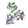
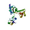
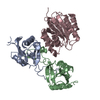
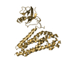
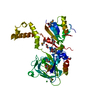
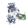
[English] 日本語
Yorodumi
- PDB-7p8d: Crystal structure of the Receiver domain of A. thaliana cytokinin... -

+
Open data


ID or keywords:

Loading...

-
Basic information

Entry
Database: PDB / ID: 7p8d
TitleCrystal structure of the Receiver domain of A. thaliana cytokinin receptor AtCRE1 in complex with Mg2+
ComponentsReceiver domain of histidine kinase 4
KeywordsSIGNALING PROTEIN / flavodoxin-like fold / cytokinin / phosphorelay
Function / homology
Function and homology information


cytokinin receptor activity / transmembrane histidine kinase cytokinin receptor activity / embryonic root morphogenesis / regulation of shoot system development / regulation of meristem development / carbohydrate homeostasis / regulation of seed germination / sulfate transport / cellular response to sucrose stimulus / protein histidine kinase binding ...cytokinin receptor activity / transmembrane histidine kinase cytokinin receptor activity / embryonic root morphogenesis / regulation of shoot system development / regulation of meristem development / carbohydrate homeostasis / regulation of seed germination / sulfate transport / cellular response to sucrose stimulus / protein histidine kinase binding / cytokinin-activated signaling pathway / protein histidine kinase activity / response to water deprivation / cellular response to phosphate starvation / osmosensory signaling pathway / myosin phosphatase activity / protein-serine/threonine phosphatase / histidine kinase / phosphorelay signal transduction system / phosphoprotein phosphatase activity / phosphorelay sensor kinase activity / defense response to bacterium / protein phosphorylation / endoplasmic reticulum membrane / protein kinase binding / enzyme binding / endoplasmic reticulum / plasma membrane
Similarity search - Function
CHASE domain superfamily / CHASE domain / CHASE domain profile. / CHASE / CHASE domain / His Kinase A (phospho-acceptor) domain / His Kinase A (phosphoacceptor) domain / Signal transduction histidine kinase, dimerisation/phosphoacceptor domain / Phosphoribosyltransferase domain / Signal transduction histidine kinase, dimerisation/phosphoacceptor domain superfamily ...CHASE domain superfamily / CHASE domain / CHASE domain profile. / CHASE / CHASE domain / His Kinase A (phospho-acceptor) domain / His Kinase A (phosphoacceptor) domain / Signal transduction histidine kinase, dimerisation/phosphoacceptor domain / Phosphoribosyltransferase domain / Signal transduction histidine kinase, dimerisation/phosphoacceptor domain superfamily / Signal transduction histidine kinase-related protein, C-terminal / Histidine kinase domain / Histidine kinase domain profile. / Response regulator receiver domain / cheY-homologous receiver domain / Signal transduction response regulator, receiver domain / Response regulatory domain profile. / CheY-like superfamily / Histidine kinase-, DNA gyrase B-, and HSP90-like ATPase / Histidine kinase-like ATPases / Histidine kinase/HSP90-like ATPase / Histidine kinase/HSP90-like ATPase superfamily
Similarity search - Domain/homology
Biological speciesArabidopsis thaliana (thale cress)
MethodX-RAY DIFFRACTION / SYNCHROTRON / MOLECULAR REPLACEMENT / Resolution: 1.7 Å
AuthorsTran, L.H. / Urbanowicz, A. / Jasinski, M. / Jaskolski, M. / Ruszkowski, M.
Funding support Poland, 1items
OrganizationGrant numberCountry
Polish National Science CentreSONATA 2018/31/D/NZ1/03630 Poland
CitationJournal: Front Plant Sci / Year: 2021
Title: 3D Domain Swapping Dimerization of the Receiver Domain of Cytokinin Receptor CRE1 From Arabidopsis thaliana and Medicago truncatula .
Authors: Tran, L.H. / Urbanowicz, A. / Jasinski, M. / Jaskolski, M. / Ruszkowski, M.
History
DepositionJul 21, 2021Deposition site: PDBE / Processing site: PDBE
Revision 1.0Oct 20, 2021Provider: repository / Type: Initial release
Revision 1.1Feb 2, 2022Group: Structure summary / Category: entity / Item: _entity.pdbx_description
Revision 1.2May 1, 2024Group: Data collection / Refinement description
Category: chem_comp_atom / chem_comp_bond / pdbx_initial_refinement_model

-
Structure visualization

Structure viewerMolecule:
MolmilJmol/JSmol

Downloads & links

-
Assembly

Deposited unit
A: Receiver domain of histidine kinase 4
B: Receiver domain of histidine kinase 4
hetero molecules


Theoretical massNumber of molelcules
Total (without water)31,9986
Polymers31,7692
Non-polymers2294
Water3,279182
1


  • Idetical with deposited unit
  • defined by author&software
  • Evidence: gel filtration
TypeNameSymmetry operationNumber
identity operation1_555x,y,z1
Buried area4300 Å2
ΔGint-60 kcal/mol
Surface area15770 Å2
MethodPISA
Unit cell
Length a, b, c (Å)88.183, 99.202, 35.623
Angle α, β, γ (deg.)90.00, 90.00, 90.00
Int Tables number18
Space group name H-MP21212

-
Components

#1: Protein Receiver domain of histidine kinase 4 / Arabidopsis histidine kinase 4 / AtHK4 / Cytokinin receptor CYTOKININ RESPONSE 1 / AtCRE1 / ...Arabidopsis histidine kinase 4 / AtHK4 / Cytokinin receptor CYTOKININ RESPONSE 1 / AtCRE1 / Cytokinin receptor CRE1 / Phosphoprotein phosphatase AHK4 / Protein AUTHENTIC HIS-KINASE 4 / Protein ROOT AS IN WOL 1 / Protein WOODEN LEG


Mass: 15884.332 Da / Num. of mol.: 2
Source method: isolated from a genetically manipulated source
Source: (gene. exp.) Arabidopsis thaliana (thale cress) / Gene: AHK4, CRE1, RAW1, WOL, At2g01830, T23K3.2
Production host: Escherichia coli 'BL21-Gold(DE3)pLysS AG' (bacteria)
References: UniProt: Q9C5U0, histidine kinase, protein-serine/threonine phosphatase
#2: Chemical ChemComp-MPD / (4S)-2-METHYL-2,4-PENTANEDIOL


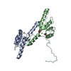
Mass: 118.174 Da / Num. of mol.: 1 / Source method: obtained synthetically / Formula: C6H14O2 / Comment: precipitant*YM
#3: Chemical ChemComp-MG / MAGNESIUM ION


Mass: 24.305 Da / Num. of mol.: 2 / Source method: obtained synthetically / Formula: Mg / Feature type: SUBJECT OF INVESTIGATION
#4: Chemical ChemComp-EDO / 1,2-ETHANEDIOL / ETHYLENE GLYCOL


Mass: 62.068 Da / Num. of mol.: 1 / Source method: obtained synthetically / Formula: C2H6O2
#5: Water ChemComp-HOH / water


Mass: 18.015 Da / Num. of mol.: 182 / Source method: isolated from a natural source / Formula: H2O
Has ligand of interestY

-
Experimental details

-
Experiment

ExperimentMethod: X-RAY DIFFRACTION / Number of used crystals: 1

-
Sample preparation

CrystalDensity Matthews: 2.62 Å3/Da / Density % sol: 52.99 %
Crystal growTemperature: 292 K / Method: vapor diffusion, sitting drop / pH: 6.5
Details: 0.03 M MgCl2, 0.03 M CaCl2 in 0.1 M MES/Imidazole at pH 6.5 and 12.5% of each PEG1000, PEG3350 and 27.5% MPD

-
Data collection

DiffractionMean temperature: 100 K / Serial crystal experiment: N
Diffraction sourceSource: SYNCHROTRON / Site: PETRA III, EMBL c/o DESY / Beamline: P13 (MX1) / Wavelength: 0.9763 Å
DetectorType: DECTRIS PILATUS 6M / Detector: PIXEL / Date: Oct 20, 2020
RadiationProtocol: SINGLE WAVELENGTH / Monochromatic (M) / Laue (L): M / Scattering type: x-ray
Radiation wavelengthWavelength: 0.9763 Å / Relative weight: 1
ReflectionResolution: 1.7→43.23 Å / Num. obs: 35267 / % possible obs: 99.9 % / Redundancy: 12.9 % / Biso Wilson estimate: 32.44 Å2 / CC1/2: 1 / Rmerge(I) obs: 0.057 / Rrim(I) all: 0.059 / Net I/σ(I): 22.9
Reflection shellResolution: 1.7→1.8 Å / Redundancy: 13.2 % / Rmerge(I) obs: 1.34 / Mean I/σ(I) obs: 1.56 / Num. unique obs: 5583 / CC1/2: 0.72 / Rrim(I) all: 1.39 / % possible all: 99.8

-
Processing

Software
NameVersionClassification
PHENIX1.18.2_3874refinement
XDSdata reduction
XDSdata scaling
PHASERphasing
RefinementMethod to determine structure: MOLECULAR REPLACEMENT
Starting model: AtCRE1-REC-K

Resolution: 1.7→43.23 Å / SU ML: 0.19 / Cross valid method: FREE R-VALUE / Phase error: 25.44 / Stereochemistry target values: ML
RfactorNum. reflection% reflection
Rfree0.2456 1058 3 %
Rwork0.1937 --
obs0.1953 35263 99.94 %
Solvent computationShrinkage radii: 0.9 Å / VDW probe radii: 1.11 Å / Solvent model: FLAT BULK SOLVENT MODEL
Refinement stepCycle: LAST / Resolution: 1.7→43.23 Å
ProteinNucleic acidLigandSolventTotal
Num. atoms2076 0 14 182 2272
Refine LS restraints
Refine-IDTypeDev idealNumber
X-RAY DIFFRACTIONf_bond_d0.012122
X-RAY DIFFRACTIONf_angle_d1.072858
X-RAY DIFFRACTIONf_dihedral_angle_d16.004801
X-RAY DIFFRACTIONf_chiral_restr0.067323
X-RAY DIFFRACTIONf_plane_restr0.007366
LS refinement shell
Resolution (Å)Rfactor RfreeNum. reflection RfreeRfactor RworkNum. reflection RworkRefine-ID% reflection obs (%)
1.7-1.780.32161290.26844186X-RAY DIFFRACTION100
1.78-1.870.28941300.24234212X-RAY DIFFRACTION100
1.87-1.990.26781310.22184219X-RAY DIFFRACTION100
1.99-2.140.2611310.20674226X-RAY DIFFRACTION100
2.14-2.360.26511310.19254242X-RAY DIFFRACTION100
2.36-2.70.24861320.20694280X-RAY DIFFRACTION100
2.7-3.40.26491340.19944326X-RAY DIFFRACTION100
3.4-43.230.22141400.17584514X-RAY DIFFRACTION100
Refinement TLS params.

Method: refined / Refine-ID: X-RAY DIFFRACTION

IDL112)L122)L132)L222)L232)L332)S11 (Å °)S12 (Å °)S13 (Å °)S21 (Å °)S22 (Å °)S23 (Å °)S31 (Å °)S32 (Å °)S33 (Å °)T112)T122)T132)T222)T232)T332)Origin x (Å)Origin y (Å)Origin z (Å)
14.86360.6394-0.13828.8642-1.05814.9822-0.1942-0.24910.3104-0.07810.42120.02-0.65380.3528-0.24650.3440.01740.03810.297-0.03640.18895.059126.666618.1098
23.1209-1.98251.03536.5315-1.75284.1449-0.0207-0.3354-0.08510.5880.07890.0404-0.106-0.045-0.03170.30360.01210.05740.33890.01730.23783.518322.078220.6167
34.3094-2.4316-0.19837.44550.18665.5471-0.1755-0.0706-0.28130.27170.37590.57990.1992-0.1076-0.17550.25830.03060.02610.27640.07110.2636-3.171322.257613.305
47.181-2.31351.49562.643-0.57290.96810.33470.3298-1.0209-0.2628-0.28481.46980.0678-0.2741-0.03350.27780.0351-0.05270.403-0.03390.4712-10.48326.56965.507
58.0927-5.86432.10827.388-2.21024.90410.3550.36490.3085-0.4376-0.3429-0.23680.17350.05970.010.2356-0.0052-0.01010.24670.01250.2164-2.161829.97027.604
65.0964-4.5009-2.03124.13452.11052.761-0.1701-1.06230.27240.89890.253-0.1076-0.1761-0.15740.05510.41480.09320.0760.50080.05070.378413.24795.1756.8046
79.28226.3503-6.19144.796-5.63838.79120.24590.55070.5154-0.41210.34030.5755-0.6544-0.4065-0.53740.40960.10950.0510.40880.01850.393220.65447.7434-2.3419
86.54041.29213.17664.20193.23183.28110.4187-0.4497-0.1060.6374-1.7819-1.29081.70492.30650.83190.76030.05460.05321.01380.3290.573319.064920.3324-19.2939
92.75751.89692.25226.01073.13284.4571-0.4346-0.00740.8795-0.19160.51330.223-1.94140.06090.04050.6241-0.08580.03770.4382-0.06680.45989.104432.3742-33.8879
109.73266.37141.50826.14223.98645.2149-0.11850.1538-1.06290.40230.0451-0.85890.22340.48860.01060.2908-0.02280.04120.34480.04880.486434.22864.3435-12.5355
118.1689-1.05462.31014.4723-1.10184.3029-0.14250.3899-0.1289-0.22280.07030.0864-0.32070.10410.07540.2785-0.0067-0.00520.2253-0.02950.20619.46754.2915-15.6987
125.3606-5.14110.40486.84570.25254.7411-0.05430.1179-0.9156-0.28210.06740.87750.4773-0.24010.0010.3272-0.04880.03940.2552-0.01580.435419.3698-9.2585-11.6979
136.1587-1.911-2.50257.85140.5926.56610.1447-0.2183-0.3481-0.0255-0.0840.8696-0.1817-0.6717-0.06690.27420.02810.00950.32560.03560.391811.6441-0.6589-6.51
146.5533-6.85330.01378.3562-0.26682.3371-0.2892-0.257-0.87590.34290.32640.81670.3709-0.0232-0.03160.36760.01920.11360.31060.06320.422321.3765-10.0299-1.3067
158.7194-4.3367-1.77285.2933-0.53294.53880.02060.057-0.1892-0.4369-0.1522-0.13620.19550.02150.06410.30610.0465-0.01810.28540.00370.25145.989518.14710.765
164.1463-0.3109-2.39624.97984.22195.4776-0.443-0.73871.49470.78810.25480.0341-0.4797-0.51320.14060.49620.06330.00060.60110.04040.76521.194111.75053.7476
Refinement TLS group
IDRefine-IDRefine TLS-IDSelection details
1X-RAY DIFFRACTION1chain 'A' and (resid 917 through 930 )
2X-RAY DIFFRACTION2chain 'A' and (resid 931 through 960 )
3X-RAY DIFFRACTION3chain 'A' and (resid 961 through 980 )
4X-RAY DIFFRACTION4chain 'A' and (resid 981 through 999 )
5X-RAY DIFFRACTION5chain 'A' and (resid 1000 through 1011 )
6X-RAY DIFFRACTION6chain 'A' and (resid 1012 through 1025 )
7X-RAY DIFFRACTION7chain 'A' and (resid 1026 through 1035 )
8X-RAY DIFFRACTION8chain 'A' and (resid 1036 through 1045 )
9X-RAY DIFFRACTION9chain 'A' and (resid 1046 through 1055 )
10X-RAY DIFFRACTION10chain 'B' and (resid 916 through 922 )
11X-RAY DIFFRACTION11chain 'B' and (resid 923 through 951 )
12X-RAY DIFFRACTION12chain 'B' and (resid 952 through 968 )
13X-RAY DIFFRACTION13chain 'B' and (resid 969 through 980 )
14X-RAY DIFFRACTION14chain 'B' and (resid 981 through 1011 )
15X-RAY DIFFRACTION15chain 'B' and (resid 1012 through 1033 )
16X-RAY DIFFRACTION16chain 'B' and (resid 1034 through 1042 )

+
About Yorodumi

-
News

-
Feb 9, 2022. New format data for meta-information of EMDB entries

New format data for meta-information of EMDB entries

  • Version 3 of the EMDB header file is now the official format.
  • The previous official version 1.9 will be removed from the archive.

Related info.:EMDB header

External links:wwPDB to switch to version 3 of the EMDB data model

-
Aug 12, 2020. Covid-19 info

Covid-19 info

URL: https://pdbjlvh1.pdbj.org/emnavi/covid19.php

New page: Covid-19 featured information page in EM Navigator.

Related info.:Covid-19 info / Mar 5, 2020. Novel coronavirus structure data

+
Mar 5, 2020. Novel coronavirus structure data

Novel coronavirus structure data

Related info.:Yorodumi Speices / Aug 12, 2020. Covid-19 info

External links:COVID-19 featured content - PDBj / Molecule of the Month (242):Coronavirus Proteases

+
Jan 31, 2019. EMDB accession codes are about to change! (news from PDBe EMDB page)

EMDB accession codes are about to change! (news from PDBe EMDB page)

  • The allocation of 4 digits for EMDB accession codes will soon come to an end. Whilst these codes will remain in use, new EMDB accession codes will include an additional digit and will expand incrementally as the available range of codes is exhausted. The current 4-digit format prefixed with “EMD-” (i.e. EMD-XXXX) will advance to a 5-digit format (i.e. EMD-XXXXX), and so on. It is currently estimated that the 4-digit codes will be depleted around Spring 2019, at which point the 5-digit format will come into force.
  • The EM Navigator/Yorodumi systems omit the EMD- prefix.

Related info.:Q: What is EMD? / ID/Accession-code notation in Yorodumi/EM Navigator

External links:EMDB Accession Codes are Changing Soon! / Contact to PDBj

+
Jul 12, 2017. Major update of PDB

Major update of PDB

  • wwPDB released updated PDB data conforming to the new PDBx/mmCIF dictionary.
  • This is a major update changing the version number from 4 to 5, and with Remediation, in which all the entries are updated.
  • In this update, many items about electron microscopy experimental information are reorganized (e.g. em_software).
  • Now, EM Navigator and Yorodumi are based on the updated data.

External links:wwPDB Remediation / Enriched Model Files Conforming to OneDep Data Standards Now Available in the PDB FTP Archive

-
Yorodumi

Thousand views of thousand structures

  • Yorodumi is a browser for structure data from EMDB, PDB, SASBDB, etc.
  • This page is also the successor to EM Navigator detail page, and also detail information page/front-end page for Omokage search.
  • The word "yorodu" (or yorozu) is an old Japanese word meaning "ten thousand". "mi" (miru) is to see.

Related info.:EMDB / PDB / SASBDB / Comparison of 3 databanks / Yorodumi Search / Aug 31, 2016. New EM Navigator & Yorodumi / Yorodumi Papers / Jmol/JSmol / Function and homology information / Changes in new EM Navigator and Yorodumi

Read more