 Movie
Movie Controller

[English] 日本語
 Yorodumi
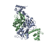
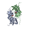
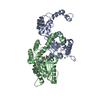
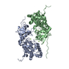
- PDB-7p47: Structure of the E3 ligase Smc5/Nse2 in complex with Ubc9-SUMO th... -
+ Open data

- Basic information
| Entry | Database: PDB / ID: 7p47 | ||||||
|---|---|---|---|---|---|---|---|
| Title | Structure of the E3 ligase Smc5/Nse2 in complex with Ubc9-SUMO thioester mimetic | ||||||
 Components Components |
| ||||||
 Keywords Keywords | LIGASE / SUMO E3 ligase activity / DNA repair | ||||||
| Function / homology |  Function and homology information Function and homology informationSmc5-Smc6 complex / SUMO conjugating enzyme activity / resolution of DNA recombination intermediates / DNA double-strand break attachment to nuclear envelope / SUMO ligase activity / mitotic spindle elongation / chromosome separation / SUMO is conjugated to E1 (UBA2:SAE1) / SUMOylation of nuclear envelope proteins / SUMO is transferred from E1 to E2 (UBE2I, UBC9) ...Smc5-Smc6 complex / SUMO conjugating enzyme activity / resolution of DNA recombination intermediates / DNA double-strand break attachment to nuclear envelope / SUMO ligase activity / mitotic spindle elongation / chromosome separation / SUMO is conjugated to E1 (UBA2:SAE1) / SUMOylation of nuclear envelope proteins / SUMO is transferred from E1 to E2 (UBE2I, UBC9) / SUMO is proteolytically processed / SUMOylation of transcription factors / Postmitotic nuclear pore complex (NPC) reformation / SUMOylation of transcription cofactors / septin ring / SUMOylation of DNA damage response and repair proteins / Transcriptional and post-translational regulation of MITF-M expression and activity / SUMOylation of DNA replication proteins / SUMOylation of SUMOylation proteins / Recruitment and ATM-mediated phosphorylation of repair and signaling proteins at DNA double strand breaks / chromatin looping / SUMOylation of RNA binding proteins / Transferases; Acyltransferases; Aminoacyltransferases / SUMO transferase activity / SUMOylation of chromatin organization proteins / recombinational repair / regulation of telomere maintenance / ubiquitin-like protein ligase binding / protein sumoylation / protein serine/threonine kinase inhibitor activity / condensed nuclear chromosome / double-strand break repair via homologous recombination / protein tag activity / nuclear envelope / single-stranded DNA binding / damaged DNA binding / chromosome, telomeric region / cell division / DNA repair / ATP hydrolysis activity / zinc ion binding / ATP binding / identical protein binding / nucleus / cytoplasm Similarity search - Function | ||||||
| Biological species |  | ||||||
| Method |  X-RAY DIFFRACTION / SYNCHROTRON / MOLECULAR REPLACEMENT / Resolution: 3.314 Å X-RAY DIFFRACTION / SYNCHROTRON / MOLECULAR REPLACEMENT / Resolution: 3.314 Å | ||||||
 Authors Authors | Lascorz, J. / Varejao, N. / Reverter, D. | ||||||
| Funding support |  Spain, 1items Spain, 1items
| ||||||
 Citation Citation | Journal: Nat Commun / Year: 2021 Title: Structural basis for the E3 ligase activity enhancement of yeast Nse2 by SUMO-interacting motifs. Authors: Varejao, N. / Lascorz, J. / Codina-Fabra, J. / Belli, G. / Borras-Gas, H. / Torres-Rosell, J. / Reverter, D. | ||||||
| History |
|
- Structure visualization
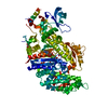
| Structure viewer | Molecule: MolmilJmol/JSmol |
|---|
- Downloads & links
-Download
| PDBx/mmCIF format | 7p47.cif.gz | 244.5 KB | Display | PDBx/mmCIF format |
|---|---|---|---|---|
| PDB format | pdb7p47.ent.gz | 195.3 KB | Display | PDB format |
| PDBx/mmJSON format | 7p47.json.gz | Tree view | PDBx/mmJSON format | |
| Others |  Other downloads Other downloads |
-Validation report
| Summary document | 7p47_validation.pdf.gz | 495.7 KB | Display | wwPDB validaton report |
|---|---|---|---|---|
| Full document | 7p47_full_validation.pdf.gz | 511 KB | Display | |
| Data in XML | 7p47_validation.xml.gz | 23 KB | Display | |
| Data in CIF | 7p47_validation.cif.gz | 31.4 KB | Display | |
| Arichive directory | https://data.pdbj.org/pub/pdb/validation_reports/p4/7p47ftp://data.pdbj.org/pub/pdb/validation_reports/p4/7p47 | HTTPS FTP |
-Related structure data
| Related structure data |  3htkS S: Starting model for refinement |
|---|---|
| Similar structure data |
-Links
 PDBj
PDBj


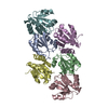
- Assembly
| Deposited unit | 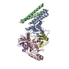
| ||||||||
|---|---|---|---|---|---|---|---|---|---|
| 1 |
| ||||||||
| Unit cell |
|
-Components
| #1: Protein | Mass: 9978.620 Da / Num. of mol.: 1 Source method: isolated from a genetically manipulated source Source: (gene. exp.) Gene: SMC5, YOL034W / Production host:  | ||||
|---|---|---|---|---|---|
| #2: Protein | Mass: 19071.645 Da / Num. of mol.: 1 Source method: isolated from a genetically manipulated source Details: Point mutants A129K, K153R Isopeptidic bond between K129 (chain A) and G98 (chain D) Source: (gene. exp.) Gene: UBC9, YDL064W / Production host: References: UniProt: P50623, Transferases; Acyltransferases; Aminoacyltransferases | ||||
| #3: Protein | Mass: 24117.096 Da / Num. of mol.: 1 Source method: isolated from a genetically manipulated source Source: (gene. exp.) Gene: MMS21, NSE2, YEL019C / Production host: References: UniProt: P38632, Transferases; Acyltransferases; Aminoacyltransferases | ||||
| #4: Protein | Mass: 13739.396 Da / Num. of mol.: 2 Source method: isolated from a genetically manipulated source Source: (gene. exp.) Gene: SMT3, YDR510W, D9719.15 / Production host: #5: Chemical | ChemComp-ZN / |   Mass: 65.409 Da / Num. of mol.: 1 / Source method: obtained synthetically / Formula: Zn / Feature type: SUBJECT OF INVESTIGATION Mass: 65.409 Da / Num. of mol.: 1 / Source method: obtained synthetically / Formula: Zn / Feature type: SUBJECT OF INVESTIGATIONHas ligand of interest | Y | |
-Experimental details
-Experiment
| Experiment | Method: X-RAY DIFFRACTION / Number of used crystals: 1 |
|---|
- Sample preparation
| Crystal | Density Matthews: 2.78 Å3/Da / Density % sol: 55.75 % |
|---|---|
| Crystal grow | Temperature: 311 K / Method: vapor diffusion, hanging drop Details: 12% PEG8000, 0.2M dimethyl-2-hydroxyethylammoniumpropane sulfonate (NDSB 211), 8% ethylene glycol, 0.1M MES pH 6.5 |
-Data collection
| Diffraction | Mean temperature: 100 K / Serial crystal experiment: N |
|---|---|
| Diffraction source | Source: SYNCHROTRON / Site: ALBA / Beamline: XALOC / Wavelength: 0.9792 Å |
| Detector | Type: DECTRIS PILATUS 6M / Detector: PIXEL / Date: May 27, 2021 |
| Radiation | Protocol: SINGLE WAVELENGTH / Monochromatic (M) / Laue (L): M / Scattering type: x-ray |
| Radiation wavelength | Wavelength: 0.9792 Å / Relative weight: 1 |
| Reflection | Resolution: 3.31→47.14 Å / Num. obs: 13097 / % possible obs: 99.2 % / Redundancy: 5.4 % / CC1/2: 0.99 / Net I/σ(I): 9.7 |
| Reflection shell | Resolution: 3.31→3.58 Å / Num. unique obs: 2585 / CC1/2: 0.68 |
- Processing
| Software |
| ||||||||||||||||||||||||||||||||||||||||
|---|---|---|---|---|---|---|---|---|---|---|---|---|---|---|---|---|---|---|---|---|---|---|---|---|---|---|---|---|---|---|---|---|---|---|---|---|---|---|---|---|---|
| Refinement | Method to determine structure: MOLECULAR REPLACEMENT Starting model: 3HTK Resolution: 3.314→47.135 Å / SU ML: 0.47 / Cross valid method: THROUGHOUT / σ(F): 1.34 / Phase error: 32.58 / Stereochemistry target values: ML
| ||||||||||||||||||||||||||||||||||||||||
| Solvent computation | Shrinkage radii: 0.9 Å / VDW probe radii: 1.11 Å / Solvent model: FLAT BULK SOLVENT MODEL | ||||||||||||||||||||||||||||||||||||||||
| Displacement parameters | Biso max: 198.58 Å2 / Biso mean: 98.82 Å2 / Biso min: 57.74 Å2 | ||||||||||||||||||||||||||||||||||||||||
| Refinement step | Cycle: final / Resolution: 3.314→47.135 Å
| ||||||||||||||||||||||||||||||||||||||||
| Refine LS restraints |
| ||||||||||||||||||||||||||||||||||||||||
| LS refinement shell | Refine-ID: X-RAY DIFFRACTION / Rfactor Rfree error: 0
| ||||||||||||||||||||||||||||||||||||||||
| Refinement TLS params. | Method: refined / Origin x: 19.2457 Å / Origin y: -12.4467 Å / Origin z: -20.4649 Å
| ||||||||||||||||||||||||||||||||||||||||
| Refinement TLS group |
|
