 Movie
Movie Controller

[English] 日本語
 Yorodumi
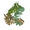
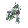
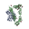
- PDB-7p3x: Homology model of the full-length AP-3 complex in a compact open ... -
+ Open data

- Basic information
| Entry | Database: PDB / ID: 7p3x | ||||||
|---|---|---|---|---|---|---|---|
| Title | Homology model of the full-length AP-3 complex in a compact open conformation | ||||||
 Components Components |
| ||||||
 Keywords Keywords | TRANSPORT PROTEIN / adaptor protein / vesicle transport / AP-3 / homology model | ||||||
| Function / homology |  Function and homology information Function and homology informationAP-3 adaptor complex / Golgi to vacuole transport / clathrin adaptor complex / membrane coat / protein targeting to vacuole / vesicle-mediated transport / cytoplasmic vesicle membrane / intracellular protein transport / endocytosis / cytoplasmic vesicle / Golgi apparatus Similarity search - Function | ||||||
| Biological species |  | ||||||
| Method | ELECTRON MICROSCOPY / single particle reconstruction / cryo EM / Resolution: 9.1 Å | ||||||
 Authors Authors | Schubert, E. / Raunser, S. | ||||||
| Funding support |  Germany, 1items Germany, 1items
| ||||||
 Citation Citation | Journal: J Biol Chem / Year: 2021 Title: Flexible open conformation of the AP-3 complex explains its role in cargo recruitment at the Golgi. Authors: Jannis Schoppe / Evelyn Schubert / Amir Apelbaum / Erdal Yavavli / Oliver Birkholz / Heike Stephanowitz / Yaping Han / Angela Perz / Oliver Hofnagel / Fan Liu / Jacob Piehler / Stefan ...Authors: Jannis Schoppe / Evelyn Schubert / Amir Apelbaum / Erdal Yavavli / Oliver Birkholz / Heike Stephanowitz / Yaping Han / Angela Perz / Oliver Hofnagel / Fan Liu / Jacob Piehler / Stefan Raunser / Christian Ungermann / Abstract: Vesicle formation at endomembranes requires the selective concentration of cargo by coat proteins. Conserved adapter protein complexes at the Golgi (AP-3), the endosome (AP-1), or the plasma membrane ...Vesicle formation at endomembranes requires the selective concentration of cargo by coat proteins. Conserved adapter protein complexes at the Golgi (AP-3), the endosome (AP-1), or the plasma membrane (AP-2) with their conserved core domain and flexible ear domains mediate this function. These complexes also rely on the small GTPase Arf1 and/or specific phosphoinositides for membrane binding. The structural details that influence these processes, however, are still poorly understood. Here we present cryo-EM structures of the full-length stable 300 kDa yeast AP-3 complex. The structures reveal that AP-3 adopts an open conformation in solution, comparable to the membrane-bound conformations of AP-1 or AP-2. This open conformation appears to be far more flexible than AP-1 or AP-2, resulting in compact, intermediate, and stretched subconformations. Mass spectrometrical analysis of the cross-linked AP-3 complex further indicates that the ear domains are flexibly attached to the surface of the complex. Using biochemical reconstitution assays, we also show that efficient AP-3 recruitment to the membrane depends primarily on cargo binding. Once bound to cargo, AP-3 clustered and immobilized cargo molecules, as revealed by single-molecule imaging on polymer-supported membranes. We conclude that its flexible open state may enable AP-3 to bind and collect cargo at the Golgi and could thus allow coordinated vesicle formation at the trans-Golgi upon Arf1 activation. | ||||||
| History |
|
- Structure visualization
| Movie |
Movie viewer |
|---|---|
| Structure viewer | Molecule: MolmilJmol/JSmol |
- Downloads & links
-Download
| PDBx/mmCIF format | 7p3x.cif.gz | 364.8 KB | Display | PDBx/mmCIF format |
|---|---|---|---|---|
| PDB format | pdb7p3x.ent.gz | 246.4 KB | Display | PDB format |
| PDBx/mmJSON format | 7p3x.json.gz | Tree view | PDBx/mmJSON format | |
| Others |  Other downloads Other downloads |
-Validation report
| Summary document | 7p3x_validation.pdf.gz | 818.2 KB | Display | wwPDB validaton report |
|---|---|---|---|---|
| Full document | 7p3x_full_validation.pdf.gz | 1.4 MB | Display | |
| Data in XML | 7p3x_validation.xml.gz | 151.5 KB | Display | |
| Data in CIF | 7p3x_validation.cif.gz | 201.7 KB | Display | |
| Arichive directory | https://data.pdbj.org/pub/pdb/validation_reports/p3/7p3xftp://data.pdbj.org/pub/pdb/validation_reports/p3/7p3x | HTTPS FTP |
-Related structure data
| Related structure data |  13187MC  7p3yC  7p3zC M: map data used to model this data C: citing same article ( |
|---|---|
| Similar structure data |


-Links
 PDBj
PDBj
- Assembly
| Deposited unit | 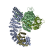
|
|---|---|
| 1 |
|
-Components
| #1: Protein | Mass: 110903.016 Da / Num. of mol.: 1 Source method: isolated from a genetically manipulated source Source: (gene. exp.) Gene: PACBIOSEQ_LOCUS5975 / Production host: |
|---|---|
| #2: Protein | Mass: 91712.016 Da / Num. of mol.: 1 Source method: isolated from a genetically manipulated source Source: (gene. exp.) Gene: PACBIOSEQ_LOCUS2762 / Production host: |
| #3: Protein | Mass: 21951.646 Da / Num. of mol.: 1 Source method: isolated from a genetically manipulated source Source: (gene. exp.) Gene: PACBIOSEQ_LOCUS3239, PACBIOSEQ_LOCUS3310, SCNYR20_0009016500, SCP684_0009016000 Production host: |
| #4: Protein | Mass: 54899.062 Da / Num. of mol.: 1 Source method: isolated from a genetically manipulated source Source: (gene. exp.) Gene: APM3, YKS6, YBR288C, YBR2035 / Production host: |
-Experimental details
-Experiment
| Experiment | Method: ELECTRON MICROSCOPY |
|---|---|
| EM experiment | Aggregation state: PARTICLE / 3D reconstruction method: single particle reconstruction |
- Sample preparation
| Component | Name: Full-length AP-3 complex from Saccharomyces cerevisiae Type: COMPLEX / Entity ID: all / Source: RECOMBINANT |
|---|---|
| Source (natural) | Organism: |
| Source (recombinant) | Organism: |
| Buffer solution | pH: 7.4 / Details: 20 mM Hepes pH 7.4 150 mM NaCl 1.5 mM MgCl2 |
| Specimen | Conc.: 0.25 mg/ml / Embedding applied: NO / Shadowing applied: NO / Staining applied: NO / Vitrification applied: YES |
| Specimen support | Grid material: GOLD / Grid mesh size: 400 divisions/in. / Grid type: UltrAuFoil R1.2/1.3 |
| Vitrification | Instrument: FEI VITROBOT MARK III / Cryogen name: ETHANE / Humidity: 100 % / Chamber temperature: 277.15 K Details: The sample was blotted using a 2.5 s blotting time and 0 blotting force with 100% humidity at 277.15 K |
- Electron microscopy imaging
| Experimental equipment |  Model: Titan Krios / Image courtesy: FEI Company |
|---|---|
| Microscopy | Model: FEI TITAN KRIOS |
| Electron gun | Electron source:  FIELD EMISSION GUN / Accelerating voltage: 300 kV / Illumination mode: SPOT SCAN FIELD EMISSION GUN / Accelerating voltage: 300 kV / Illumination mode: SPOT SCAN |
| Electron lens | Mode: BRIGHT FIELD / Nominal magnification: 130000 X / Nominal defocus max: 3600 nm / Nominal defocus min: 1500 nm |
| Specimen holder | Specimen holder model: FEI TITAN KRIOS AUTOGRID HOLDER |
| Image recording | Electron dose: 81 e/Å2 / Detector mode: COUNTING / Film or detector model: GATAN K2 SUMMIT (4k x 4k) |
- Processing
| EM software |
| ||||||||||||||||||||||||||||||||||||||||
|---|---|---|---|---|---|---|---|---|---|---|---|---|---|---|---|---|---|---|---|---|---|---|---|---|---|---|---|---|---|---|---|---|---|---|---|---|---|---|---|---|---|
| CTF correction | Type: PHASE FLIPPING ONLY | ||||||||||||||||||||||||||||||||||||||||
| Particle selection | Num. of particles selected: 958892 | ||||||||||||||||||||||||||||||||||||||||
| Symmetry | Point symmetry: C1 (asymmetric) | ||||||||||||||||||||||||||||||||||||||||
| 3D reconstruction | Resolution: 9.1 Å / Resolution method: FSC 0.5 CUT-OFF / Num. of particles: 23039 / Symmetry type: POINT | ||||||||||||||||||||||||||||||||||||||||
| Atomic model building | Protocol: FLEXIBLE FIT |
