 Movie
Movie Controller

[English] 日本語
 Yorodumi
- PDB-7p2e: Human mitochondrial ribosome small subunit in complex with IF3, G... -
+ Open data

- Basic information
| Entry | Database: PDB / ID: 7p2e | ||||||||||||
|---|---|---|---|---|---|---|---|---|---|---|---|---|---|
| Title | Human mitochondrial ribosome small subunit in complex with IF3, GMPPMP and streptomycin | ||||||||||||
 Components Components |
| ||||||||||||
 Keywords Keywords | RIBOSOME / Initiation factor 3 / translation / antibiotic / streptomycin | ||||||||||||
| Function / homology |  Function and homology information Function and homology informationmitochondrial translational initiation / translation factor activity, RNA binding / mitochondrial ribosome assembly / Mitochondrial translation elongation / Mitochondrial translation initiation / Mitochondrial ribosome-associated quality control / Mitochondrial translation termination / mitochondrial ribosome / ribosome disassembly / mitochondrial small ribosomal subunit ...mitochondrial translational initiation / translation factor activity, RNA binding / mitochondrial ribosome assembly / Mitochondrial translation elongation / Mitochondrial translation initiation / Mitochondrial ribosome-associated quality control / Mitochondrial translation termination / mitochondrial ribosome / ribosome disassembly / mitochondrial small ribosomal subunit / sperm head-tail coupling apparatus / mitochondrial translation / apoptotic mitochondrial changes / positive regulation of proteolysis / ribosomal small subunit binding / translation initiation factor activity / Mitochondrial protein degradation / apoptotic signaling pathway / fibrillar center / cell junction / regulation of translation / ribosome binding / small ribosomal subunit / nuclear membrane / small ribosomal subunit rRNA binding / Hydrolases; Acting on acid anhydrides; Acting on GTP to facilitate cellular and subcellular movement / cell population proliferation / tRNA binding / mitochondrial inner membrane / rRNA binding / structural constituent of ribosome / ribosome / translation / mitochondrial matrix / protein domain specific binding / mRNA binding / GTPase activity / nucleolus / GTP binding / mitochondrion / RNA binding / nucleoplasm / nucleus / plasma membrane / cytosol / cytoplasm Similarity search - Function | ||||||||||||
| Biological species |  Homo sapiens (human) Homo sapiens (human) | ||||||||||||
| Method | ELECTRON MICROSCOPY / single particle reconstruction / cryo EM / Resolution: 2.4 Å | ||||||||||||
 Authors Authors | Itoh, Y. / Khawaja, A. / Singh, V. / Rorbach, J. / Amunts, A. | ||||||||||||
| Funding support |  Sweden, 2items Sweden, 2items
| ||||||||||||
 Citation Citation | Journal: Elife / Year: 2022 Title: Structure of the mitoribosomal small subunit with streptomycin reveals Fe-S clusters and physiological molecules. Authors: Yuzuru Itoh / Vivek Singh / Anas Khawaja / Andreas Naschberger / Minh Duc Nguyen / Joanna Rorbach / Alexey Amunts / Abstract: The mitoribosome regulates cellular energy production, and its dysfunction is associated with aging. Inhibition of the mitoribosome can be caused by off-target binding of antimicrobial drugs and was ...The mitoribosome regulates cellular energy production, and its dysfunction is associated with aging. Inhibition of the mitoribosome can be caused by off-target binding of antimicrobial drugs and was shown to be coupled with a bilateral decreased visual acuity. Previously, we reported mitochondria-specific protein aspects of the mitoribosome, and in this article we present a 2.4-Å resolution structure of the small subunit in a complex with the anti-tuberculosis drug streptomycin that reveals roles of non-protein components. We found iron-sulfur clusters that are coordinated by different mitoribosomal proteins, nicotinamide adenine dinucleotide (NAD) associated with rRNA insertion, and posttranslational modifications. This is the first evidence of inter-protein coordination of iron-sulfur, and the finding of iron-sulfur clusters and NAD as fundamental building blocks of the mitoribosome directly links to mitochondrial disease and aging. We also report details of streptomycin interactions, suggesting that the mitoribosome-bound streptomycin is likely to be in hydrated gem-diol form and can be subjected to other modifications by the cellular milieu. The presented approach of adding antibiotics to cultured cells can be used to define their native structures in a bound form under more physiological conditions, and since streptomycin is a widely used drug for treatment, the newly resolved features can serve as determinants for targeting. | ||||||||||||
| History |
|
- Structure visualization
| Structure viewer | Molecule: MolmilJmol/JSmol |
|---|
- Downloads & links
-Download
| PDBx/mmCIF format | 7p2e.cif.gz | 3.1 MB | Display | PDBx/mmCIF format |
|---|---|---|---|---|
| PDB format | pdb7p2e.ent.gz | Display | PDB format | |
| PDBx/mmJSON format | 7p2e.json.gz | Tree view | PDBx/mmJSON format | |
| Others |  Other downloads Other downloads |
-Validation report
| Arichive directory | https://data.pdbj.org/pub/pdb/validation_reports/p2/7p2eftp://data.pdbj.org/pub/pdb/validation_reports/p2/7p2e | HTTPS FTP |
|---|
-Related structure data
| Related structure data |  13170MC M: map data used to model this data C: citing same article ( |
|---|---|
| Similar structure data |
-Links
 PDBj
PDBj


- Assembly
| Deposited unit | 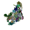
|
|---|---|
| 1 |
|
-Components
-RNA chain , 1 types, 1 molecules A
| #1: RNA chain | Mass: 306547.531 Da / Num. of mol.: 1 / Source method: isolated from a natural source / Details: cell / Source: (natural) Homo sapiens (human) |
|---|
+28S ribosomal protein ... , 27 types, 27 molecules BCDEFGHIJKLMNOPQRSTUVWXYZ01
-Protein , 4 types, 4 molecules 2348
| #29: Protein | Mass: 13409.661 Da / Num. of mol.: 1 / Source method: isolated from a natural source / Details: cell / Source: (natural) Homo sapiens (human) / References: UniProt: Q96BP2 |
|---|---|
| #30: Protein | Mass: 22395.326 Da / Num. of mol.: 1 / Source method: isolated from a natural source / Details: cell / Source: (natural) Homo sapiens (human) / References: UniProt: Q9NWT8 |
| #31: Protein | Mass: 78648.547 Da / Num. of mol.: 1 / Source method: isolated from a natural source / Details: cell / Source: (natural) Homo sapiens (human) / References: UniProt: Q96EY7 |
| #32: Protein | Mass: 32545.461 Da / Num. of mol.: 1 Source method: isolated from a genetically manipulated source Details: Natural variant Thr68Ile and Phe263Leu / Source: (gene. exp.) Homo sapiens (human) / Description: cell / Gene: MTIF3, DC38 / Production host: Homo sapiens (human) / References: UniProt: Q9H2K0 |
-Non-polymers , 10 types, 3176 molecules 


| #33: Chemical | ChemComp-NAD /  Mass: 663.425 Da / Num. of mol.: 1 / Source method: obtained synthetically / Formula: C21H27N7O14P2 / Feature type: SUBJECT OF INVESTIGATION / Comment: NAD*YM Mass: 663.425 Da / Num. of mol.: 1 / Source method: obtained synthetically / Formula: C21H27N7O14P2 / Feature type: SUBJECT OF INVESTIGATION / Comment: NAD*YM | ||||||||||||
|---|---|---|---|---|---|---|---|---|---|---|---|---|---|
| #34: Chemical | ChemComp-SPM /  Mass: 202.340 Da / Num. of mol.: 1 / Source method: obtained synthetically / Formula: C10H26N4 / Feature type: SUBJECT OF INVESTIGATION Mass: 202.340 Da / Num. of mol.: 1 / Source method: obtained synthetically / Formula: C10H26N4 / Feature type: SUBJECT OF INVESTIGATION | ||||||||||||
| #35: Chemical | ChemComp-5I0 / [( Mass: 602.613 Da / Num. of mol.: 1 / Source method: obtained synthetically / Formula: C21H44N7O13 / Feature type: SUBJECT OF INVESTIGATION Mass: 602.613 Da / Num. of mol.: 1 / Source method: obtained synthetically / Formula: C21H44N7O13 / Feature type: SUBJECT OF INVESTIGATION | ||||||||||||
| #36: Chemical | ChemComp-K /  Mass: 39.098 Da / Num. of mol.: 21 / Source method: obtained synthetically / Formula: K Mass: 39.098 Da / Num. of mol.: 21 / Source method: obtained synthetically / Formula: K#37: Chemical | ChemComp-MG /  Mass: 24.305 Da / Num. of mol.: 62 / Source method: obtained synthetically / Formula: Mg Mass: 24.305 Da / Num. of mol.: 62 / Source method: obtained synthetically / Formula: Mg#38: Chemical | ChemComp-ZN / |  Mass: 65.409 Da / Num. of mol.: 1 / Source method: obtained synthetically / Formula: Zn Mass: 65.409 Da / Num. of mol.: 1 / Source method: obtained synthetically / Formula: Zn#39: Chemical |  Mass: 175.820 Da / Num. of mol.: 2 / Source method: obtained synthetically / Formula: Fe2S2 / Feature type: SUBJECT OF INVESTIGATION Mass: 175.820 Da / Num. of mol.: 2 / Source method: obtained synthetically / Formula: Fe2S2 / Feature type: SUBJECT OF INVESTIGATION#40: Chemical | ChemComp-ATP / |  Mass: 507.181 Da / Num. of mol.: 1 / Source method: obtained synthetically / Formula: C10H16N5O13P3 / Feature type: SUBJECT OF INVESTIGATION / Comment: ATP, energy-carrying molecule*YM Mass: 507.181 Da / Num. of mol.: 1 / Source method: obtained synthetically / Formula: C10H16N5O13P3 / Feature type: SUBJECT OF INVESTIGATION / Comment: ATP, energy-carrying molecule*YM#41: Chemical | ChemComp-GNP / |  Mass: 522.196 Da / Num. of mol.: 1 / Source method: obtained synthetically / Formula: C10H17N6O13P3 / Feature type: SUBJECT OF INVESTIGATION Mass: 522.196 Da / Num. of mol.: 1 / Source method: obtained synthetically / Formula: C10H17N6O13P3 / Feature type: SUBJECT OF INVESTIGATIONComment: GppNHp, GMPPNP, energy-carrying molecule analogue*YM #42: Water | ChemComp-HOH / | Mass: 18.015 Da / Num. of mol.: 3085 / Source method: isolated from a natural source / Formula: H2O |
-Details
| Has ligand of interest | Y |
|---|
-Experimental details
-Experiment
| Experiment | Method: ELECTRON MICROSCOPY |
|---|---|
| EM experiment | Aggregation state: PARTICLE / 3D reconstruction method: single particle reconstruction |
- Sample preparation
| Component |
| ||||||||||||||||||||||||||||||
|---|---|---|---|---|---|---|---|---|---|---|---|---|---|---|---|---|---|---|---|---|---|---|---|---|---|---|---|---|---|---|---|
| Molecular weight | Experimental value: NO | ||||||||||||||||||||||||||||||
| Source (natural) |
| ||||||||||||||||||||||||||||||
| Source (recombinant) | Organism: Homo sapiens (human) | ||||||||||||||||||||||||||||||
| Buffer solution | pH: 7.5 | ||||||||||||||||||||||||||||||
| Buffer component |
| ||||||||||||||||||||||||||||||
| Specimen | Embedding applied: NO / Shadowing applied: NO / Staining applied: NO / Vitrification applied: YES | ||||||||||||||||||||||||||||||
| Specimen support | Grid material: COPPER / Grid type: Quantifoil R2/1 | ||||||||||||||||||||||||||||||
| Vitrification | Instrument: FEI VITROBOT MARK IV / Cryogen name: ETHANE / Humidity: 100 % / Chamber temperature: 297 K |
- Electron microscopy imaging
| Experimental equipment |  Model: Titan Krios / Image courtesy: FEI Company |
|---|---|
| Microscopy | Model: FEI TITAN KRIOS |
| Electron gun | Electron source:  FIELD EMISSION GUN / Accelerating voltage: 300 kV / Illumination mode: FLOOD BEAM FIELD EMISSION GUN / Accelerating voltage: 300 kV / Illumination mode: FLOOD BEAM |
| Electron lens | Mode: BRIGHT FIELD / Nominal defocus max: 4000 nm / Nominal defocus min: 500 nm / Cs: 2.7 mm / C2 aperture diameter: 70 µm |
| Specimen holder | Cryogen: NITROGEN / Specimen holder model: FEI TITAN KRIOS AUTOGRID HOLDER |
| Image recording | Average exposure time: 4 sec. / Electron dose: 30 e/Å2 / Detector mode: COUNTING / Film or detector model: GATAN K2 SUMMIT (4k x 4k) / Num. of real images: 31773 |
| EM imaging optics | Energyfilter name: GIF Quantum LS / Energyfilter slit width: 20 eV |
| Image scans | Movie frames/image: 20 / Used frames/image: 1-20 |
- Processing
| EM software |
| ||||||||||||||||||||||||||||||||||||
|---|---|---|---|---|---|---|---|---|---|---|---|---|---|---|---|---|---|---|---|---|---|---|---|---|---|---|---|---|---|---|---|---|---|---|---|---|---|
| CTF correction | Type: PHASE FLIPPING AND AMPLITUDE CORRECTION | ||||||||||||||||||||||||||||||||||||
| Particle selection | Num. of particles selected: 2487123 | ||||||||||||||||||||||||||||||||||||
| Symmetry | Point symmetry: C1 (asymmetric) | ||||||||||||||||||||||||||||||||||||
| 3D reconstruction | Resolution: 2.4 Å / Resolution method: FSC 0.143 CUT-OFF / Num. of particles: 885199 / Algorithm: BACK PROJECTION / Symmetry type: POINT | ||||||||||||||||||||||||||||||||||||
| Atomic model building | Space: REAL | ||||||||||||||||||||||||||||||||||||
| Atomic model building | PDB-ID: 6RW4 Accession code: 6RW4 / Source name: PDB / Type: experimental model |

