 Movie
Movie Controller

[English] 日本語
 Yorodumi
- PDB-7p20: High resolution structure of the Juniperus ashei allergen - Jun a 3 -
+ Open data

- Basic information
| Entry | Database: PDB / ID: 7p20 | ||||||
|---|---|---|---|---|---|---|---|
| Title | High resolution structure of the Juniperus ashei allergen - Jun a 3 | ||||||
 Components Components | Pathogenesis-related 5 protein Jun a 3.0101 | ||||||
 Keywords Keywords | ALLERGEN / Thaumatin-like protein / Juniperus Ashei | ||||||
| Function / homology |  Function and homology information Function and homology information | ||||||
| Biological species |  Juniperus ashei (Ozark white cedar) Juniperus ashei (Ozark white cedar) | ||||||
| Method |  X-RAY DIFFRACTION / SYNCHROTRON / MOLECULAR REPLACEMENT / Resolution: 1.4 Å X-RAY DIFFRACTION / SYNCHROTRON / MOLECULAR REPLACEMENT / Resolution: 1.4 Å | ||||||
 Authors Authors | Eder, M. / Hofer, G. / Keller, W. | ||||||
| Funding support |  Austria, 1items Austria, 1items
| ||||||
 Citation Citation | Journal: To Be Published Title: Crystal structure of the allergen Jun a 3 the Thaumatin-like protein of Juniperus ashei Authors: Eder, M. / Hofer, G. / Keller, W. | ||||||
| History |
|
- Structure visualization
| Structure viewer | Molecule: MolmilJmol/JSmol |
|---|
- Downloads & links
-Download
| PDBx/mmCIF format | 7p20.cif.gz | 104.1 KB | Display | PDBx/mmCIF format |
|---|---|---|---|---|
| PDB format | pdb7p20.ent.gz | 77.2 KB | Display | PDB format |
| PDBx/mmJSON format | 7p20.json.gz | Tree view | PDBx/mmJSON format | |
| Others |  Other downloads Other downloads |
-Validation report
| Arichive directory | https://data.pdbj.org/pub/pdb/validation_reports/p2/7p20ftp://data.pdbj.org/pub/pdb/validation_reports/p2/7p20 | HTTPS FTP |
|---|
-Related structure data
| Related structure data | 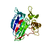 2ahnS S: Starting model for refinement |
|---|---|
| Similar structure data |
-Links
 PDBj
PDBj
- Assembly
| Deposited unit | 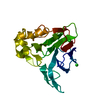
| |||||||||
|---|---|---|---|---|---|---|---|---|---|---|
| 1 |
| |||||||||
| Unit cell |
| |||||||||
| Components on special symmetry positions |
|
-Components
| #1: Protein | Mass: 22021.402 Da / Num. of mol.: 1 / Mutation: N133Q, N188Q Source method: isolated from a genetically manipulated source Source: (gene. exp.) Juniperus ashei (Ozark white cedar) / Plasmid: pcDNA3.4 / Cell line (production host): Expi293F / Production host:  Homo sapiens (human) / Tissue (production host): Kidney (Embryonic) / References: UniProt: P81295 Homo sapiens (human) / Tissue (production host): Kidney (Embryonic) / References: UniProt: P81295 | ||||||
|---|---|---|---|---|---|---|---|
| #2: Chemical |   Mass: 35.453 Da / Num. of mol.: 2 / Source method: isolated from a natural source / Formula: Cl Mass: 35.453 Da / Num. of mol.: 2 / Source method: isolated from a natural source / Formula: Cl#3: Water | ChemComp-HOH / |  Mass: 18.015 Da / Num. of mol.: 290 / Source method: isolated from a natural source / Formula: H2O Mass: 18.015 Da / Num. of mol.: 290 / Source method: isolated from a natural source / Formula: H2OHas ligand of interest | N | Has protein modification | Y | |
-Experimental details
-Experiment
| Experiment | Method: X-RAY DIFFRACTION / Number of used crystals: 1 |
|---|
- Sample preparation
| Crystal | Density Matthews: 2.03 Å3/Da / Density % sol: 39.31 % / Description: big leaflets |
|---|---|
| Crystal grow | Temperature: 293 K / Method: microbatch / pH: 4.5 Details: Index condition F5 (0.1 M Ammonium acetate, 0.1 M BIS-TRIS pH 5.5, and 17% w/v Polyethylene glycol 10,000) |
-Data collection
| Diffraction | Mean temperature: 100 K / Serial crystal experiment: N |
|---|---|
| Diffraction source | Source: SYNCHROTRON / Site: ESRF  / Beamline: ID29 / Wavelength: 1.2543 Å / Beamline: ID29 / Wavelength: 1.2543 Å |
| Detector | Type: DECTRIS PILATUS 6M / Detector: PIXEL / Date: Oct 3, 2018 |
| Radiation | Protocol: SINGLE WAVELENGTH / Monochromatic (M) / Laue (L): M / Scattering type: x-ray |
| Radiation wavelength | Wavelength: 1.2543 Å / Relative weight: 1 |
| Reflection | Resolution: 1.4→35.34 Å / Num. obs: 34665 / % possible obs: 99.68 % / Redundancy: 6.2 % / CC1/2: 0.99 / Rmerge(I) obs: 0.09554 / Rpim(I) all: 0.04089 / Rrim(I) all: 0.1041 / Net I/σ(I): 18.44 |
| Reflection shell | Resolution: 1.4→1.45 Å / Redundancy: 5.2 % / Rmerge(I) obs: 0.1447 / Mean I/σ(I) obs: 11.95 / Num. unique obs: 3393 / CC1/2: 0.984 / Rpim(I) all: 0.06866 / Rrim(I) all: 0.1608 / % possible all: 97.67 |
- Processing
| Software |
| |||||||||||||||||||||||||||||||||||||||||||||||||||||||||||||||
|---|---|---|---|---|---|---|---|---|---|---|---|---|---|---|---|---|---|---|---|---|---|---|---|---|---|---|---|---|---|---|---|---|---|---|---|---|---|---|---|---|---|---|---|---|---|---|---|---|---|---|---|---|---|---|---|---|---|---|---|---|---|---|---|---|
| Refinement | Method to determine structure: MOLECULAR REPLACEMENT Starting model: 2ahn Resolution: 1.4→35.34 Å / SU ML: 0.09 / Cross valid method: THROUGHOUT / σ(F): 1.41 / Phase error: 16.17 / Stereochemistry target values: ML
| |||||||||||||||||||||||||||||||||||||||||||||||||||||||||||||||
| Solvent computation | Shrinkage radii: 0.9 Å / VDW probe radii: 1.11 Å / Solvent model: FLAT BULK SOLVENT MODEL | |||||||||||||||||||||||||||||||||||||||||||||||||||||||||||||||
| Displacement parameters | Biso max: 84.15 Å2 / Biso mean: 17.2939 Å2 / Biso min: 5.54 Å2 | |||||||||||||||||||||||||||||||||||||||||||||||||||||||||||||||
| Refinement step | Cycle: final / Resolution: 1.4→35.34 Å
| |||||||||||||||||||||||||||||||||||||||||||||||||||||||||||||||
| LS refinement shell | Refine-ID: X-RAY DIFFRACTION / Rfactor Rfree error: 0 / Total num. of bins used: 8
|
