 Movie
Movie Controller

[English] 日本語
 Yorodumi
- PDB-7otl: Structure of a psychrophilic CCA-adding enzyme crystallized by co... -
+ Open data

- Basic information
| Entry | Database: PDB / ID: 7otl | |||||||||
|---|---|---|---|---|---|---|---|---|---|---|
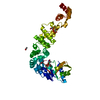
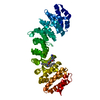
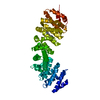
| Title | Structure of a psychrophilic CCA-adding enzyme crystallized by counter-diffusion | |||||||||
 Components Components | CCA-adding enzyme | |||||||||
 Keywords Keywords | RNA BINDING PROTEIN / tRNA maturation / tRNA nucleotidyltransferase / psychrophilic enzyme | |||||||||
| Function / homology |  Function and homology information Function and homology informationtRNA processing / nucleotidyltransferase activity / tRNA binding / nucleotide binding / metal ion binding Similarity search - Function | |||||||||
| Biological species |  Planococcus halocryophilus (bacteria) Planococcus halocryophilus (bacteria) | |||||||||
| Method |  X-RAY DIFFRACTION / SYNCHROTRON / MOLECULAR REPLACEMENT / Resolution: 2.8 Å X-RAY DIFFRACTION / SYNCHROTRON / MOLECULAR REPLACEMENT / Resolution: 2.8 Å | |||||||||
 Authors Authors | Rollet, K. / de Wijn, R. / Hennig, O. / Betat, H. / Moerl, M. / Lorber, B. / Sauter, C. | |||||||||
| Funding support |  France, 2items France, 2items
| |||||||||
 Citation Citation | Journal: Comput Struct Biotechnol J / Year: 2021 Title: CCA-addition in the cold: Structural characterization of the psychrophilic CCA-adding enzyme from the permafrost bacterium Planococcus halocryophilus . Authors: de Wijn, R. / Rollet, K. / Ernst, F.G.M. / Wellner, K. / Betat, H. / Morl, M. / Sauter, C. | |||||||||
| History |
|
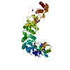
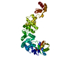
- Structure visualization
| Structure viewer | Molecule: MolmilJmol/JSmol |
|---|
- Downloads & links
-Download
| PDBx/mmCIF format | 7otl.cif.gz | 166 KB | Display | PDBx/mmCIF format |
|---|---|---|---|---|
| PDB format | pdb7otl.ent.gz | 124.1 KB | Display | PDB format |
| PDBx/mmJSON format | 7otl.json.gz | Tree view | PDBx/mmJSON format | |
| Others |  Other downloads Other downloads |
-Validation report
| Arichive directory | https://data.pdbj.org/pub/pdb/validation_reports/ot/7otlftp://data.pdbj.org/pub/pdb/validation_reports/ot/7otl | HTTPS FTP |
|---|
-Related structure data
| Related structure data |  7oqxC  7otrC 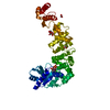 6qxnS S: Starting model for refinement C: citing same article ( |
|---|---|
| Similar structure data |

-Links
 PDBj
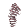
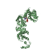
PDBj- Assembly
| Deposited unit | 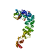
| ||||||||||||
|---|---|---|---|---|---|---|---|---|---|---|---|---|---|
| 1 |
| ||||||||||||
| Unit cell |
|
-Components
| #1: Protein | Mass: 48498.266 Da / Num. of mol.: 1 Source method: isolated from a genetically manipulated source Source: (gene. exp.) Planococcus halocryophilus (bacteria) / Gene: BBI08_05760 / Plasmid: pET-30b(+) / Details (production host): pET-30bEk/LIC / Production host: | ||||
|---|---|---|---|---|---|
| #2: Chemical | ChemComp-SO4 /   Mass: 96.063 Da / Num. of mol.: 4 / Source method: obtained synthetically / Formula: SO4 Mass: 96.063 Da / Num. of mol.: 4 / Source method: obtained synthetically / Formula: SO4#3: Water | ChemComp-HOH / |  Mass: 18.015 Da / Num. of mol.: 20 / Source method: isolated from a natural source / Formula: H2O Mass: 18.015 Da / Num. of mol.: 20 / Source method: isolated from a natural source / Formula: H2OHas ligand of interest | N | |
-Experimental details
-Experiment
| Experiment | Method: X-RAY DIFFRACTION / Number of used crystals: 1 |
|---|
- Sample preparation
| Crystal | Density Matthews: 3.7 Å3/Da / Density % sol: 66.72 % / Description: bipyramid |
|---|---|
| Crystal grow | Temperature: 293 K / Method: counter-diffusion / pH: 5 Details: Protein solution at 4.5 mg/mL in 50 mM Tris-HCl pH 7.5, 200 mM NaCl, 5 mM MgCl2; Crystals were obtained in capillaries filled with protein solution and sealed at one extremity and plunged in ...Details: Protein solution at 4.5 mg/mL in 50 mM Tris-HCl pH 7.5, 200 mM NaCl, 5 mM MgCl2; Crystals were obtained in capillaries filled with protein solution and sealed at one extremity and plunged in 3 M ammonium sulfate, 0.1 M sodium acetate pH 5. |
-Data collection
| Diffraction | Mean temperature: 100 K / Serial crystal experiment: N |
|---|---|
| Diffraction source | Source: SYNCHROTRON / Site: SLS  / Beamline: X06DA / Wavelength: 1 Å / Beamline: X06DA / Wavelength: 1 Å |
| Detector | Type: DECTRIS PILATUS 2M-F / Detector: PIXEL / Date: Jun 26, 2016 |
| Radiation | Protocol: SINGLE WAVELENGTH / Monochromatic (M) / Laue (L): M / Scattering type: x-ray |
| Radiation wavelength | Wavelength: 1 Å / Relative weight: 1 |
| Reflection | Resolution: 2.8→50 Å / Num. obs: 18615 / % possible obs: 98.6 % / Redundancy: 55.7 % / Biso Wilson estimate: 60.29 Å2 / CC1/2: 0.999 / Rrim(I) all: 0.258 / Net I/σ(I): 21.3 |
| Reflection shell | Resolution: 2.8→2.9 Å / Redundancy: 57.5 % / Mean I/σ(I) obs: 3.1 / Num. unique obs: 1819 / CC1/2: 1 / Rrim(I) all: 2.04 / % possible all: 100 |
- Processing
| Software |
| |||||||||||||||||||||||||||||||||||||||||||||||||
|---|---|---|---|---|---|---|---|---|---|---|---|---|---|---|---|---|---|---|---|---|---|---|---|---|---|---|---|---|---|---|---|---|---|---|---|---|---|---|---|---|---|---|---|---|---|---|---|---|---|---|
| Refinement | Method to determine structure: MOLECULAR REPLACEMENT Starting model: 6QXN Resolution: 2.8→46.88 Å / SU ML: 0.3942 / Cross valid method: FREE R-VALUE / σ(F): 1.34 / Phase error: 25.3979 Stereochemistry target values: GeoStd + Monomer Library + CDL v1.2
| |||||||||||||||||||||||||||||||||||||||||||||||||
| Solvent computation | Shrinkage radii: 0.9 Å / VDW probe radii: 1.11 Å / Solvent model: FLAT BULK SOLVENT MODEL | |||||||||||||||||||||||||||||||||||||||||||||||||
| Displacement parameters | Biso mean: 53.31 Å2 | |||||||||||||||||||||||||||||||||||||||||||||||||
| Refinement step | Cycle: LAST / Resolution: 2.8→46.88 Å
| |||||||||||||||||||||||||||||||||||||||||||||||||
| Refine LS restraints |
| |||||||||||||||||||||||||||||||||||||||||||||||||
| LS refinement shell |
|
