 Movie
Movie Controller

[English] 日本語
 Yorodumi
- PDB-7oso: The crystal structure of Erwinia tasmaniensis levansucrase in com... -
+ Open data

- Basic information
| Entry | Database: PDB / ID: 7oso | ||||||
|---|---|---|---|---|---|---|---|
| Title | The crystal structure of Erwinia tasmaniensis levansucrase in complex with (S)-1,2,4-butanentriol | ||||||
 Components Components | Levansucrase (Beta-D-fructofuranosyl transferase) | ||||||
 Keywords Keywords | TRANSFERASE / fructosyltransferase / levan / FOS / transfructosylation / fructo-oligosaccharides / oligofructans | ||||||
| Function / homology | levansucrase / levansucrase activity / Glycoside hydrolase, family 68 / Levansucrase/Invertase / carbohydrate utilization / Glycosyl hydrolase, five-bladed beta-propellor domain superfamily / metal ion binding / (2~{S})-butane-1,2,4-triol / levansucrase Function and homology information Function and homology information | ||||||
| Biological species |  Erwinia tasmaniensis (bacteria) Erwinia tasmaniensis (bacteria) | ||||||
| Method |  X-RAY DIFFRACTION / SYNCHROTRON / MOLECULAR REPLACEMENT / molecular replacement / Resolution: 1.4 Å X-RAY DIFFRACTION / SYNCHROTRON / MOLECULAR REPLACEMENT / molecular replacement / Resolution: 1.4 Å | ||||||
 Authors Authors | Polsinelli, I. / Salomone-Stagni, M. / Benini, S. | ||||||
 Citation Citation | Journal: Acta Crystallogr.,Sect.F / Year: 2022 Title: Erwinia tasmaniensis levansucrase shows enantiomer selection for (S)-1,2,4-butanetriol. Authors: Polsinelli, I. / Salomone-Stagni, M. / Benini, S. #1: Journal: Int J Mol Sci / Year: 2019Title: The Structure of Sucrose-Soaked Levansucrase Crystals from Erwinia tasmaniensis reveals a Binding Pocket for Levanbiose. Authors: Polsinelli, I. / Caliandro, R. / Demitri, N. / Benini, S. #2: Journal: Int J Biol Macromol / Year: 2019Title: Comparison of the Levansucrase from the epiphyte Erwinia tasmaniensis vs its homologue from the phytopathogen Erwinia amylovora. Authors: Polsinelli, I. / Caliandro, R. / Salomone-Stagni, M. / Demitri, N. / Rejzek, M. / Field, R.A. / Benini, S. | ||||||
| History |
|
- Structure visualization
| Structure viewer | Molecule: MolmilJmol/JSmol |
|---|
- Downloads & links
-Download
| PDBx/mmCIF format | 7oso.cif.gz | 202.8 KB | Display | PDBx/mmCIF format |
|---|---|---|---|---|
| PDB format | pdb7oso.ent.gz | 158.1 KB | Display | PDB format |
| PDBx/mmJSON format | 7oso.json.gz | Tree view | PDBx/mmJSON format | |
| Others |  Other downloads Other downloads |
-Validation report
| Summary document | 7oso_validation.pdf.gz | 747.1 KB | Display | wwPDB validaton report |
|---|---|---|---|---|
| Full document | 7oso_full_validation.pdf.gz | 747.3 KB | Display | |
| Data in XML | 7oso_validation.xml.gz | 21.4 KB | Display | |
| Data in CIF | 7oso_validation.cif.gz | 33.9 KB | Display | |
| Arichive directory | https://data.pdbj.org/pub/pdb/validation_reports/os/7osoftp://data.pdbj.org/pub/pdb/validation_reports/os/7oso | HTTPS FTP |
-Related structure data
| Related structure data | 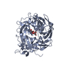 4d47S S: Starting model for refinement |
|---|---|
| Similar structure data |
-Links
 PDBj
PDBj- Assembly
| Deposited unit | 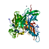
| ||||||||
|---|---|---|---|---|---|---|---|---|---|
| 1 |
| ||||||||
| Unit cell |
|
-Components
| #1: Protein | Mass: 46139.234 Da / Num. of mol.: 1 Source method: isolated from a genetically manipulated source Source: (gene. exp.) Erwinia tasmaniensis (strain DSM 17950 / CIP 109463 / Et1/99) (bacteria)Strain: DSM 17950 / CIP 109463 / Et1/99 / Gene: lsc, ETA_34670 Production host: References: UniProt: B2VCC3, levansucrase | ||||||
|---|---|---|---|---|---|---|---|
| #2: Chemical |   Mass: 65.409 Da / Num. of mol.: 3 / Source method: obtained synthetically / Formula: Zn Mass: 65.409 Da / Num. of mol.: 3 / Source method: obtained synthetically / Formula: Zn#3: Chemical | ChemComp-0V1 / ( |   Mass: 106.120 Da / Num. of mol.: 1 / Source method: obtained synthetically / Formula: C4H10O3 / Feature type: SUBJECT OF INVESTIGATION Mass: 106.120 Da / Num. of mol.: 1 / Source method: obtained synthetically / Formula: C4H10O3 / Feature type: SUBJECT OF INVESTIGATION#4: Water | ChemComp-HOH / |  Mass: 18.015 Da / Num. of mol.: 517 / Source method: isolated from a natural source / Formula: H2O Mass: 18.015 Da / Num. of mol.: 517 / Source method: isolated from a natural source / Formula: H2OHas ligand of interest | Y | |
-Experimental details
-Experiment
| Experiment | Method: X-RAY DIFFRACTION / Number of used crystals: 1 |
|---|
- Sample preparation
| Crystal | Density Matthews: 2.7 Å3/Da / Density % sol: 54.38 % |
|---|---|
| Crystal grow | Temperature: 293 K / Method: vapor diffusion, hanging drop Details: PEG 3000 15%, 1,2,4-Butanetriol (CAS 3068-00-6) 20%, NDSB 256 1%, 2.5 mM Manganese(II) chloride tetrahydrate, 2.5 mM Cobalt(II) chloride hexahydrate, 2.5 mM Nickel(II) chloride hexahydrate, ...Details: PEG 3000 15%, 1,2,4-Butanetriol (CAS 3068-00-6) 20%, NDSB 256 1%, 2.5 mM Manganese(II) chloride tetrahydrate, 2.5 mM Cobalt(II) chloride hexahydrate, 2.5 mM Nickel(II) chloride hexahydrate, 2.5 mM Zinc acetate dihydrate) |
-Data collection
| Diffraction | Mean temperature: 100 K / Serial crystal experiment: N |
|---|---|
| Diffraction source | Source: SYNCHROTRON / Site: ELETTRA  / Beamline: 5.2R / Wavelength: 1 Å / Beamline: 5.2R / Wavelength: 1 Å |
| Detector | Type: DECTRIS PILATUS3 2M / Detector: PIXEL / Date: Jul 20, 2017 |
| Radiation | Protocol: SINGLE WAVELENGTH / Monochromatic (M) / Laue (L): M / Scattering type: x-ray |
| Radiation wavelength | Wavelength: 1 Å / Relative weight: 1 |
| Reflection | Resolution: 1.4→45.19 Å / Num. obs: 189742 / % possible obs: 99.91 % / Redundancy: 5.6 % / Biso Wilson estimate: 20.95 Å2 / CC1/2: 1 / Rmerge(I) obs: 0.05 / Net I/σ(I): 23.25 |
| Reflection shell | Resolution: 1.4→1.45 Å / Num. unique obs: 30398 / CC1/2: 0.686 |
-Phasing
| Phasing | Method: molecular replacement |
|---|
- Processing
| Software |
| |||||||||||||||||||||||||||||||||||||||||||||||||||||||||||||||||
|---|---|---|---|---|---|---|---|---|---|---|---|---|---|---|---|---|---|---|---|---|---|---|---|---|---|---|---|---|---|---|---|---|---|---|---|---|---|---|---|---|---|---|---|---|---|---|---|---|---|---|---|---|---|---|---|---|---|---|---|---|---|---|---|---|---|---|
| Refinement | Method to determine structure: MOLECULAR REPLACEMENT Starting model: 4D47 Resolution: 1.4→45.19 Å / Cor.coef. Fo:Fc: 0.98 / Cor.coef. Fo:Fc free: 0.975 / SU B: 2.117 / SU ML: 0.035 / SU R Cruickshank DPI: 0.0474 / Cross valid method: THROUGHOUT / σ(F): 0 / ESU R: 0.047 / ESU R Free: 0.047 / Stereochemistry target values: MAXIMUM LIKELIHOOD Details: HYDROGENS HAVE BEEN ADDED IN THE RIDING POSITIONS U VALUES : REFINED INDIVIDUALLY
| |||||||||||||||||||||||||||||||||||||||||||||||||||||||||||||||||
| Solvent computation | Ion probe radii: 0.8 Å / Shrinkage radii: 0.8 Å / VDW probe radii: 1.2 Å / Solvent model: MASK | |||||||||||||||||||||||||||||||||||||||||||||||||||||||||||||||||
| Displacement parameters | Biso max: 109.49 Å2 / Biso mean: 24.355 Å2 / Biso min: 15.31 Å2
| |||||||||||||||||||||||||||||||||||||||||||||||||||||||||||||||||
| Refinement step | Cycle: final / Resolution: 1.4→45.19 Å
| |||||||||||||||||||||||||||||||||||||||||||||||||||||||||||||||||
| Refine LS restraints |
| |||||||||||||||||||||||||||||||||||||||||||||||||||||||||||||||||
| LS refinement shell | Resolution: 1.4→1.436 Å / Rfactor Rfree error: 0
|
