 Movie
Movie Controller

[English] 日本語
 Yorodumi
- PDB-7op9: Purine nucleoside phosphorylase(DeoD-type) from H. pylori with 2,... -
+ Open data

- Basic information
| Entry | Database: PDB / ID: 7op9 | ||||||||||||
|---|---|---|---|---|---|---|---|---|---|---|---|---|---|
| Title | Purine nucleoside phosphorylase(DeoD-type) from H. pylori with 2,6-dichloropurine | ||||||||||||
 Components Components | Purine nucleoside phosphorylase DeoD-type | ||||||||||||
 Keywords Keywords | TRANSFERASE / inhibitor / complex | ||||||||||||
| Function / homology |  Function and homology information Function and homology informationpurine-nucleoside phosphorylase / purine-nucleoside phosphorylase activity / purine nucleoside catabolic process / cytosol Similarity search - Function | ||||||||||||
| Biological species |   Helicobacter pylori (bacteria) Helicobacter pylori (bacteria) | ||||||||||||
| Method |  X-RAY DIFFRACTION / SYNCHROTRON / MOLECULAR REPLACEMENT / Resolution: 1.5 Å X-RAY DIFFRACTION / SYNCHROTRON / MOLECULAR REPLACEMENT / Resolution: 1.5 Å | ||||||||||||
 Authors Authors | Narczyk, M. / Stefanic, Z. | ||||||||||||
| Funding support |  Poland, Poland,  Croatia, 3items Croatia, 3items
| ||||||||||||
 Citation Citation | Journal: J Enzyme Inhib Med Chem / Year: 2022 Title: Interactions of 2,6-substituted purines with purine nucleoside phosphorylase from Helicobacter pylori in solution and in the crystal, and the effects of these compounds on cell cultures of this bacterium. Authors: Narczyk, M. / Wojtys, M.I. / Lescic Asler, I. / Zinic, B. / Luic, M. / Jagusztyn-Krynicka, E.K. / Stefanic, Z. / Bzowska, A. | ||||||||||||
| History |
|
- Structure visualization
| Structure viewer | Molecule: MolmilJmol/JSmol |
|---|
- Downloads & links
-Download
| PDBx/mmCIF format | 7op9.cif.gz | 595.8 KB | Display | PDBx/mmCIF format |
|---|---|---|---|---|
| PDB format | pdb7op9.ent.gz | 486.9 KB | Display | PDB format |
| PDBx/mmJSON format | 7op9.json.gz | Tree view | PDBx/mmJSON format | |
| Others |  Other downloads Other downloads |
-Validation report
| Summary document | 7op9_validation.pdf.gz | 4.4 MB | Display | wwPDB validaton report |
|---|---|---|---|---|
| Full document | 7op9_full_validation.pdf.gz | 4.5 MB | Display | |
| Data in XML | 7op9_validation.xml.gz | 129.3 KB | Display | |
| Data in CIF | 7op9_validation.cif.gz | 183 KB | Display | |
| Arichive directory | https://data.pdbj.org/pub/pdb/validation_reports/op/7op9ftp://data.pdbj.org/pub/pdb/validation_reports/op/7op9 | HTTPS FTP |
-Related structure data
| Related structure data |  7ooyC  7oozC  7opaC  6f4xS C: citing same article ( S: Starting model for refinement |
|---|---|
| Similar structure data |
-Links
 PDBj
PDBj


- Assembly
| Deposited unit | 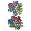
| ||||||||
|---|---|---|---|---|---|---|---|---|---|
| 1 | 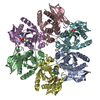
| ||||||||
| 2 | 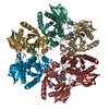
| ||||||||
| Unit cell |
|
-Components
| #1: Protein | Mass: 25866.104 Da / Num. of mol.: 12 Source method: isolated from a genetically manipulated source Source: (gene. exp.) Helicobacter pylori (strain ATCC 700392 / 26695) (bacteria)Strain: ATCC 700392 / 26695 / Gene: deoD, HP_1178 / Production host: References: UniProt: P56463, purine-nucleoside phosphorylase #2: Chemical | ChemComp-06K / 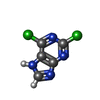  Mass: 189.002 Da / Num. of mol.: 12 / Source method: obtained synthetically / Formula: C5H2Cl2N4 / Feature type: SUBJECT OF INVESTIGATION Mass: 189.002 Da / Num. of mol.: 12 / Source method: obtained synthetically / Formula: C5H2Cl2N4 / Feature type: SUBJECT OF INVESTIGATION#3: Chemical | ChemComp-IMD /   Mass: 69.085 Da / Num. of mol.: 13 / Source method: obtained synthetically / Formula: C3H5N2 Mass: 69.085 Da / Num. of mol.: 13 / Source method: obtained synthetically / Formula: C3H5N2#4: Chemical | ChemComp-MG /   Mass: 24.305 Da / Num. of mol.: 12 / Source method: obtained synthetically / Formula: Mg Mass: 24.305 Da / Num. of mol.: 12 / Source method: obtained synthetically / Formula: Mg#5: Water | ChemComp-HOH / |  Mass: 18.015 Da / Num. of mol.: 2661 / Source method: isolated from a natural source / Formula: H2O Mass: 18.015 Da / Num. of mol.: 2661 / Source method: isolated from a natural source / Formula: H2OHas ligand of interest | Y | |
|---|
-Experimental details
-Experiment
| Experiment | Method: X-RAY DIFFRACTION / Number of used crystals: 1 |
|---|
- Sample preparation
| Crystal | Density Matthews: 2.28 Å3/Da / Density % sol: 46.12 % |
|---|---|
| Crystal grow | Temperature: 294.15 K / Method: vapor diffusion, hanging drop / pH: 7 / Details: 0.2 M imidazole pH 7,0, PPG 400 |
-Data collection
| Diffraction | Mean temperature: 100 K / Serial crystal experiment: N | ||||||||||||||||||||||||||||||||||||||||||||||||||||||||||||||||||||||||||||||||||||||||||
|---|---|---|---|---|---|---|---|---|---|---|---|---|---|---|---|---|---|---|---|---|---|---|---|---|---|---|---|---|---|---|---|---|---|---|---|---|---|---|---|---|---|---|---|---|---|---|---|---|---|---|---|---|---|---|---|---|---|---|---|---|---|---|---|---|---|---|---|---|---|---|---|---|---|---|---|---|---|---|---|---|---|---|---|---|---|---|---|---|---|---|---|
| Diffraction source | Source: SYNCHROTRON / Site: ELETTRA  / Beamline: 5.2R / Wavelength: 1 Å / Beamline: 5.2R / Wavelength: 1 Å | ||||||||||||||||||||||||||||||||||||||||||||||||||||||||||||||||||||||||||||||||||||||||||
| Detector | Type: DECTRIS PILATUS 2M / Detector: PIXEL / Date: Jan 30, 2018 | ||||||||||||||||||||||||||||||||||||||||||||||||||||||||||||||||||||||||||||||||||||||||||
| Radiation | Protocol: SINGLE WAVELENGTH / Monochromatic (M) / Laue (L): M / Scattering type: x-ray | ||||||||||||||||||||||||||||||||||||||||||||||||||||||||||||||||||||||||||||||||||||||||||
| Radiation wavelength | Wavelength: 1 Å / Relative weight: 1 | ||||||||||||||||||||||||||||||||||||||||||||||||||||||||||||||||||||||||||||||||||||||||||
| Reflection | Resolution: 1.5→46.831 Å / Num. obs: 773378 / % possible obs: 87.8 % / Observed criterion σ(I): -3 / Redundancy: 2.6 % / Biso Wilson estimate: 13.36 Å2 / CC1/2: 0.994 / Rmerge(I) obs: 0.061 / Rrim(I) all: 0.087 / Χ2: 1.021 / Net I/σ(I): 6.59 | ||||||||||||||||||||||||||||||||||||||||||||||||||||||||||||||||||||||||||||||||||||||||||
| Reflection shell | Diffraction-ID: 1 / Rejects: _
|
- Processing
| Software |
| |||||||||||||||||||||||||||||||||||||||||||||||||||||||||||||||||||||||||||||||||||||||||||||||||||||||||
|---|---|---|---|---|---|---|---|---|---|---|---|---|---|---|---|---|---|---|---|---|---|---|---|---|---|---|---|---|---|---|---|---|---|---|---|---|---|---|---|---|---|---|---|---|---|---|---|---|---|---|---|---|---|---|---|---|---|---|---|---|---|---|---|---|---|---|---|---|---|---|---|---|---|---|---|---|---|---|---|---|---|---|---|---|---|---|---|---|---|---|---|---|---|---|---|---|---|---|---|---|---|---|---|---|---|---|
| Refinement | Method to determine structure: MOLECULAR REPLACEMENT Starting model: 6F4X Resolution: 1.5→42.59 Å / SU ML: 0.18 / Cross valid method: FREE R-VALUE / σ(F): 1.96 / Phase error: 22.7 / Stereochemistry target values: ML
| |||||||||||||||||||||||||||||||||||||||||||||||||||||||||||||||||||||||||||||||||||||||||||||||||||||||||
| Solvent computation | Shrinkage radii: 0.9 Å / VDW probe radii: 1.11 Å / Solvent model: FLAT BULK SOLVENT MODEL | |||||||||||||||||||||||||||||||||||||||||||||||||||||||||||||||||||||||||||||||||||||||||||||||||||||||||
| Displacement parameters | Biso max: 67.72 Å2 / Biso mean: 19.0956 Å2 / Biso min: 4.8 Å2 | |||||||||||||||||||||||||||||||||||||||||||||||||||||||||||||||||||||||||||||||||||||||||||||||||||||||||
| Refinement step | Cycle: final / Resolution: 1.5→42.59 Å
| |||||||||||||||||||||||||||||||||||||||||||||||||||||||||||||||||||||||||||||||||||||||||||||||||||||||||
| LS refinement shell | Refine-ID: X-RAY DIFFRACTION / Total num. of bins used: 14
|
