 Movie
Movie Controller

[English] 日本語
 Yorodumi
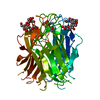
- PDB-7olu: Structure of the N-terminal domain of BC2L-C lectin (1-131) in co... -
+ Open data

- Basic information
| Entry | Database: PDB / ID: 7olu | ||||||
|---|---|---|---|---|---|---|---|
| Title | Structure of the N-terminal domain of BC2L-C lectin (1-131) in complex with a synthetic beta-C-fucoside ligand | ||||||
 Components Components | Lectin | ||||||
 Keywords Keywords | SUGAR BINDING PROTEIN / Bacterial lectin / Fucosides / Glycomimetic / Inhibitor / Anti-microbial | ||||||
| Function / homology | Lectin Bc2l-C, N-terminal / : / Bc2l-C, N-terminal domain / Calcium-mediated lectin / Calcium-mediated lectin superfamily / Fucose-binding lectin II (PA-IIL) / identical protein binding / Chem-VJT / Lectin Function and homology information Function and homology information | ||||||
| Biological species |  Burkholderia cenocepacia (bacteria) Burkholderia cenocepacia (bacteria) | ||||||
| Method |  X-RAY DIFFRACTION / SYNCHROTRON / MOLECULAR REPLACEMENT / Resolution: 1.793 Å X-RAY DIFFRACTION / SYNCHROTRON / MOLECULAR REPLACEMENT / Resolution: 1.793 Å | ||||||
 Authors Authors | Bermeo, R. / Varrot, A. | ||||||
| Funding support |  France, 1items France, 1items
| ||||||
 Citation Citation | Journal: Acs Chem.Biol. / Year: 2022 Title: Targeting a Multidrug-Resistant Pathogen: First Generation Antagonists of Burkholderia cenocepacia 's BC2L-C Lectin. Authors: Bermeo, R. / Lal, K. / Ruggeri, D. / Lanaro, D. / Mazzotta, S. / Vasile, F. / Imberty, A. / Belvisi, L. / Varrot, A. / Bernardi, A. | ||||||
| History |
|
- Structure visualization
| Structure viewer | Molecule: MolmilJmol/JSmol |
|---|
- Downloads & links
-Download
| PDBx/mmCIF format | 7olu.cif.gz | 44.8 KB | Display | PDBx/mmCIF format |
|---|---|---|---|---|
| PDB format | pdb7olu.ent.gz | Display | PDB format | |
| PDBx/mmJSON format | 7olu.json.gz | Tree view | PDBx/mmJSON format | |
| Others |  Other downloads Other downloads |
-Validation report
| Summary document | 7olu_validation.pdf.gz | 747.9 KB | Display | wwPDB validaton report |
|---|---|---|---|---|
| Full document | 7olu_full_validation.pdf.gz | 748 KB | Display | |
| Data in XML | 7olu_validation.xml.gz | 8.4 KB | Display | |
| Data in CIF | 7olu_validation.cif.gz | 11.3 KB | Display | |
| Arichive directory | https://data.pdbj.org/pub/pdb/validation_reports/ol/7oluftp://data.pdbj.org/pub/pdb/validation_reports/ol/7olu | HTTPS FTP |
-Related structure data
| Related structure data |  7olwC 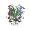 2wq4S S: Starting model for refinement C: citing same article ( |
|---|---|
| Similar structure data |


-Links
 PDBj
PDBj- Assembly
| Deposited unit | 
| |||||||||||||||
|---|---|---|---|---|---|---|---|---|---|---|---|---|---|---|---|---|
| 1 | 
| |||||||||||||||
| Unit cell |
| |||||||||||||||
| Components on special symmetry positions |
|
-Components
| #1: Protein | Mass: 14010.936 Da / Num. of mol.: 1 Source method: isolated from a genetically manipulated source Source: (gene. exp.) Burkholderia cenocepacia (strain ATCC BAA-245 / DSM 16553 / LMG 16656 / NCTC 13227 / J2315 / CF5610) (bacteria)Strain: ATCC BAA-245 / DSM 16553 / LMG 16656 / NCTC 13227 / J2315 / CF5610 Gene: BCAM0185 / Production host: |
|---|---|
| #2: Chemical | ChemComp-VJT / (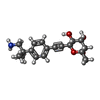  Mass: 319.395 Da / Num. of mol.: 1 / Source method: obtained synthetically / Formula: C18H25NO4 / Feature type: SUBJECT OF INVESTIGATION Mass: 319.395 Da / Num. of mol.: 1 / Source method: obtained synthetically / Formula: C18H25NO4 / Feature type: SUBJECT OF INVESTIGATION |
| #3: Water | ChemComp-HOH /  Mass: 18.015 Da / Num. of mol.: 99 / Source method: isolated from a natural source / Formula: H2O Mass: 18.015 Da / Num. of mol.: 99 / Source method: isolated from a natural source / Formula: H2O |
| Has ligand of interest | Y |
-Experimental details
-Experiment
| Experiment | Method: X-RAY DIFFRACTION / Number of used crystals: 1 |
|---|
- Sample preparation
| Crystal | Density Matthews: 1.87 Å3/Da / Density % sol: 34.35 % |
|---|---|
| Crystal grow | Temperature: 292 K / Method: vapor diffusion, hanging drop / pH: 7 / Details: sodium citrate 1.2M |
-Data collection
| Diffraction | Mean temperature: 100 K / Serial crystal experiment: N |
|---|---|
| Diffraction source | Source: SYNCHROTRON / Site: SOLEIL / Beamline: PROXIMA 2 / Wavelength: 0.98 Å |
| Detector | Type: DECTRIS EIGER X 9M / Detector: PIXEL / Date: Sep 5, 2020 |
| Radiation | Protocol: SINGLE WAVELENGTH / Monochromatic (M) / Laue (L): M / Scattering type: x-ray |
| Radiation wavelength | Wavelength: 0.98 Å / Relative weight: 1 |
| Reflection | Resolution: 1.79→47.07 Å / Num. obs: 9696 / % possible obs: 99.7 % / Redundancy: 20.2 % / Biso Wilson estimate: 20.9 Å2 / CC1/2: 1 / Rmerge(I) obs: 0.039 / Rpim(I) all: 0.013 / Rrim(I) all: 0.041 / Net I/σ(I): 49.1 |
| Reflection shell | Resolution: 1.79→1.83 Å / Redundancy: 17.2 % / Rmerge(I) obs: 0.228 / Mean I/σ(I) obs: 11.1 / Num. unique obs: 563 / CC1/2: 0.99 / Rpim(I) all: 0.077 / Rrim(I) all: 0.241 / % possible all: 95.4 |
- Processing
| Software |
| ||||||||||||||||||||||||||||||||||||||||||||||||||||||||||||||||||||||||||||||||||||||||||||||||||||||||||||||||||||||||||||||||||||||||||||||||||||||
|---|---|---|---|---|---|---|---|---|---|---|---|---|---|---|---|---|---|---|---|---|---|---|---|---|---|---|---|---|---|---|---|---|---|---|---|---|---|---|---|---|---|---|---|---|---|---|---|---|---|---|---|---|---|---|---|---|---|---|---|---|---|---|---|---|---|---|---|---|---|---|---|---|---|---|---|---|---|---|---|---|---|---|---|---|---|---|---|---|---|---|---|---|---|---|---|---|---|---|---|---|---|---|---|---|---|---|---|---|---|---|---|---|---|---|---|---|---|---|---|---|---|---|---|---|---|---|---|---|---|---|---|---|---|---|---|---|---|---|---|---|---|---|---|---|---|---|---|---|---|---|---|
| Refinement | Method to determine structure: MOLECULAR REPLACEMENT Starting model: 2wq4 Resolution: 1.793→38.065 Å / Cor.coef. Fo:Fc: 0.977 / Cor.coef. Fo:Fc free: 0.961 / SU B: 2.736 / SU ML: 0.085 / Cross valid method: FREE R-VALUE / ESU R: 0.026 / ESU R Free: 0.026 Details: Hydrogens have been added in their riding positions
| ||||||||||||||||||||||||||||||||||||||||||||||||||||||||||||||||||||||||||||||||||||||||||||||||||||||||||||||||||||||||||||||||||||||||||||||||||||||
| Solvent computation | Ion probe radii: 0.8 Å / Shrinkage radii: 0.8 Å / VDW probe radii: 1.2 Å / Solvent model: MASK BULK SOLVENT | ||||||||||||||||||||||||||||||||||||||||||||||||||||||||||||||||||||||||||||||||||||||||||||||||||||||||||||||||||||||||||||||||||||||||||||||||||||||
| Displacement parameters | Biso mean: 25.797 Å2
| ||||||||||||||||||||||||||||||||||||||||||||||||||||||||||||||||||||||||||||||||||||||||||||||||||||||||||||||||||||||||||||||||||||||||||||||||||||||
| Refinement step | Cycle: LAST / Resolution: 1.793→38.065 Å
| ||||||||||||||||||||||||||||||||||||||||||||||||||||||||||||||||||||||||||||||||||||||||||||||||||||||||||||||||||||||||||||||||||||||||||||||||||||||
| Refine LS restraints |
| ||||||||||||||||||||||||||||||||||||||||||||||||||||||||||||||||||||||||||||||||||||||||||||||||||||||||||||||||||||||||||||||||||||||||||||||||||||||
| LS refinement shell |
|
