 Movie
Movie Controller

[English] 日本語
 Yorodumi
- PDB-7ol1: The X-ray structure of L-threonine dehydrogenase from the common ... -
+ Open data

- Basic information
| Entry | Database: PDB / ID: 7ol1 | ||||||
|---|---|---|---|---|---|---|---|
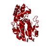
| Title | The X-ray structure of L-threonine dehydrogenase from the common hospital pathogen Clostridium difficile. | ||||||
 Components Components | L-threonine 3-dehydrogenase | ||||||
 Keywords Keywords | OXIDOREDUCTASE / Dehydrogenase / apoenzyme. | ||||||
| Function / homology | L-threonine 3-dehydrogenase / : / L-threonine 3-dehydrogenase activity / L-threonine catabolic process / NAD-dependent epimerase/dehydratase / NAD dependent epimerase/dehydratase family / NAD(P)-binding domain superfamily / L-threonine 3-dehydrogenase Function and homology information Function and homology information | ||||||
| Biological species |  Clostridioides difficile (bacteria) Clostridioides difficile (bacteria) | ||||||
| Method |  X-RAY DIFFRACTION / SYNCHROTRON / MOLECULAR REPLACEMENT / molecular replacement / Resolution: 2.6 Å X-RAY DIFFRACTION / SYNCHROTRON / MOLECULAR REPLACEMENT / molecular replacement / Resolution: 2.6 Å | ||||||
 Authors Authors | Guo, J. / Cooper, J.B. | ||||||
 Citation Citation | Journal: Acta Crystallogr.,Sect.F / Year: 2021 Title: The X-ray structure of L-threonine dehydrogenase from the common hospital pathogen Clostridium difficile. Authors: Adjogatse, E. / Bennett, J. / Guo, J. / Erskine, P.T. / Wood, S.P. / Wren, B.W. / Cooper, J.B. | ||||||
| History |
|
- Structure visualization
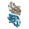
| Structure viewer | Molecule: MolmilJmol/JSmol |
|---|
- Downloads & links
-Download
| PDBx/mmCIF format | 7ol1.cif.gz | 141.7 KB | Display | PDBx/mmCIF format |
|---|---|---|---|---|
| PDB format | pdb7ol1.ent.gz | 109.9 KB | Display | PDB format |
| PDBx/mmJSON format | 7ol1.json.gz | Tree view | PDBx/mmJSON format | |
| Others |  Other downloads Other downloads |
-Validation report
| Arichive directory | https://data.pdbj.org/pub/pdb/validation_reports/ol/7ol1ftp://data.pdbj.org/pub/pdb/validation_reports/ol/7ol1 | HTTPS FTP |
|---|
-Related structure data
| Related structure data |  5lc1S S: Starting model for refinement |
|---|---|
| Similar structure data | |
| Experimental dataset #1 | Data reference: 10.5281/zenodo.4771118 / Data set type: diffraction image data |


-Links
 PDBj
PDBj- Assembly
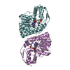
| Deposited unit | 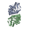
| ||||||||
|---|---|---|---|---|---|---|---|---|---|
| 1 |
| ||||||||
| Unit cell |
|
-Components
| #1: Protein | Mass: 38208.352 Da / Num. of mol.: 2 Source method: isolated from a genetically manipulated source Details: The N-terminal sequence MGHHHHHHHHHHSSGHIEGRH originates from the expression tag. Source: (gene. exp.) Clostridioides difficile (bacteria)Gene: cdgr_08060, NCTC13307_02322, SAMEA1402406_00083, SAMEA3375037_00176 Production host: References: UniProt: A0A346VYB9, L-threonine 3-dehydrogenase #2: Water | ChemComp-HOH / |  Mass: 18.015 Da / Num. of mol.: 112 / Source method: isolated from a natural source / Formula: H2O Mass: 18.015 Da / Num. of mol.: 112 / Source method: isolated from a natural source / Formula: H2O |
|---|
-Experimental details
-Experiment
| Experiment | Method: X-RAY DIFFRACTION / Number of used crystals: 1 |
|---|
- Sample preparation
| Crystal | Density Matthews: 5.8 Å3/Da / Density % sol: 78.9 % / Description: Multifaceted. |
|---|---|
| Crystal grow | Temperature: 293 K / Method: vapor diffusion, hanging drop / pH: 8.5 Details: 5 microlitres of enzyme at a concentration of 5 mg/ml and 5 microlitres of 0.2 M lithium sulphate, 0.1 M Tris-HCl pH 8.5, 30 % v/v PEG 4000 (Molecular Dimensions Limited Structure Screen I, condition 35). Temp details: Ambient temperature |
-Data collection
| Diffraction | Mean temperature: 100 K / Serial crystal experiment: N | ||||||||||||||||||||||||||||||||||||||||||||||||||||||||||||||||||||||||||||||||||||||||||||||||||||||||||||||
|---|---|---|---|---|---|---|---|---|---|---|---|---|---|---|---|---|---|---|---|---|---|---|---|---|---|---|---|---|---|---|---|---|---|---|---|---|---|---|---|---|---|---|---|---|---|---|---|---|---|---|---|---|---|---|---|---|---|---|---|---|---|---|---|---|---|---|---|---|---|---|---|---|---|---|---|---|---|---|---|---|---|---|---|---|---|---|---|---|---|---|---|---|---|---|---|---|---|---|---|---|---|---|---|---|---|---|---|---|---|---|---|
| Diffraction source | Source: SYNCHROTRON / Site: Diamond  / Beamline: I04-1 / Wavelength: 0.92 Å / Beamline: I04-1 / Wavelength: 0.92 Å | ||||||||||||||||||||||||||||||||||||||||||||||||||||||||||||||||||||||||||||||||||||||||||||||||||||||||||||||
| Detector | Type: DECTRIS PILATUS 2M / Detector: PIXEL / Date: Oct 6, 2012 | ||||||||||||||||||||||||||||||||||||||||||||||||||||||||||||||||||||||||||||||||||||||||||||||||||||||||||||||
| Radiation | Protocol: SINGLE WAVELENGTH / Monochromatic (M) / Laue (L): M / Scattering type: x-ray | ||||||||||||||||||||||||||||||||||||||||||||||||||||||||||||||||||||||||||||||||||||||||||||||||||||||||||||||
| Radiation wavelength | Wavelength: 0.92 Å / Relative weight: 1 | ||||||||||||||||||||||||||||||||||||||||||||||||||||||||||||||||||||||||||||||||||||||||||||||||||||||||||||||
| Reflection | Resolution: 2.56→63.28 Å / Num. obs: 53475 / % possible obs: 99.5 % / Redundancy: 9.3 % / CC1/2: 0.998 / Rmerge(I) obs: 0.118 / Rpim(I) all: 0.04 / Rrim(I) all: 0.125 / Rsym value: 0.118 / Net I/av σ(I): 4.2 / Net I/σ(I): 12.2 | ||||||||||||||||||||||||||||||||||||||||||||||||||||||||||||||||||||||||||||||||||||||||||||||||||||||||||||||
| Reflection shell | Diffraction-ID: 1
|
-Phasing
| Phasing | Method: molecular replacement |
|---|
- Processing
| Software |
| ||||||||||||||||||||||||||||||||||||||||||||||||||||||||||||
|---|---|---|---|---|---|---|---|---|---|---|---|---|---|---|---|---|---|---|---|---|---|---|---|---|---|---|---|---|---|---|---|---|---|---|---|---|---|---|---|---|---|---|---|---|---|---|---|---|---|---|---|---|---|---|---|---|---|---|---|---|---|
| Refinement | Method to determine structure: MOLECULAR REPLACEMENT Starting model: 5lc1 Resolution: 2.6→63.28 Å / Cor.coef. Fo:Fc: 0.958 / Cor.coef. Fo:Fc free: 0.939 / SU B: 10.429 / SU ML: 0.198 / SU R Cruickshank DPI: 0.234 / Cross valid method: FREE R-VALUE / σ(F): 0 / ESU R: 0.234 / ESU R Free: 0.21 / Stereochemistry target values: MAXIMUM LIKELIHOOD Details: HYDROGENS HAVE BEEN ADDED IN THE RIDING POSITIONS U VALUES : REFINED INDIVIDUALLY
| ||||||||||||||||||||||||||||||||||||||||||||||||||||||||||||
| Solvent computation | Ion probe radii: 0.8 Å / Shrinkage radii: 0.8 Å / VDW probe radii: 1.2 Å / Solvent model: MASK | ||||||||||||||||||||||||||||||||||||||||||||||||||||||||||||
| Displacement parameters | Biso max: 205.04 Å2 / Biso mean: 75.367 Å2 / Biso min: 27.12 Å2
| ||||||||||||||||||||||||||||||||||||||||||||||||||||||||||||
| Refinement step | Cycle: final / Resolution: 2.6→63.28 Å
| ||||||||||||||||||||||||||||||||||||||||||||||||||||||||||||
| Refine LS restraints |
| ||||||||||||||||||||||||||||||||||||||||||||||||||||||||||||
| LS refinement shell | Resolution: 2.6→2.668 Å / Rfactor Rfree error: 0 / Total num. of bins used: 20
|
