Movie
Movie Controller


+ Open data

- Basic information
| Entry | Database: PDB / ID: 7oim | |||||||||||||||
|---|---|---|---|---|---|---|---|---|---|---|---|---|---|---|---|---|
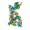
| Title | Mouse RNF213, with mixed nucleotides bound | |||||||||||||||
 Components Components | E3 ubiquitin-protein ligase RNF213 | |||||||||||||||
 Keywords Keywords | SIGNALING PROTEIN / nucleotide / AAA / RNF213 / E3 ligase | |||||||||||||||
| Function / homology |  Function and homology information Function and homology informationlipid ubiquitination / negative regulation of non-canonical Wnt signaling pathway / lipid droplet formation / xenophagy / sprouting angiogenesis / Antigen processing: Ubiquitination & Proteasome degradation / Transferases; Acyltransferases; Aminoacyltransferases / regulation of lipid metabolic process / immune system process / protein K63-linked ubiquitination ...lipid ubiquitination / negative regulation of non-canonical Wnt signaling pathway / lipid droplet formation / xenophagy / sprouting angiogenesis / Antigen processing: Ubiquitination & Proteasome degradation / Transferases; Acyltransferases; Aminoacyltransferases / regulation of lipid metabolic process / immune system process / protein K63-linked ubiquitination / protein autoubiquitination / lipid droplet / RING-type E3 ubiquitin transferase / Hydrolases; Acting on acid anhydrides; Acting on acid anhydrides to facilitate cellular and subcellular movement / ubiquitin-protein transferase activity / ubiquitin protein ligase activity / angiogenesis / ubiquitin-dependent protein catabolic process / defense response to bacterium / protein ubiquitination / nucleolus / ATP hydrolysis activity / zinc ion binding / ATP binding / cytoplasm / cytosol Similarity search - Function | |||||||||||||||
| Biological species |  | |||||||||||||||
| Method | ELECTRON MICROSCOPY / single particle reconstruction / cryo EM / Resolution: 4 Å | |||||||||||||||
 Authors Authors | Grabarczyk, D. / Ahel, J. / Clausen, T. | |||||||||||||||
| Funding support |  Austria, Austria,  United Kingdom, 4items United Kingdom, 4items
| |||||||||||||||
 Citation Citation | Journal: To Be Published Title: E3 ubiquitin ligase RNF213 employs a non-canonical zinc finger active site and is allosterically regulated by ATP Authors: Ahel, J. / Fletcher, A.J. / Grabarczyk, D. / Roitinger, E. / Deszcz, L. / Lehner, A. / Virdee, S. / Clausen, T. | |||||||||||||||
| History |
|
- Structure visualization
| Structure viewer | Molecule: MolmilJmol/JSmol |
|---|
- Downloads & links
-Download
| PDBx/mmCIF format | 7oim.cif.gz | 811.5 KB | Display | PDBx/mmCIF format |
|---|---|---|---|---|
| PDB format | pdb7oim.ent.gz | 642 KB | Display | PDB format |
| PDBx/mmJSON format | 7oim.json.gz | Tree view | PDBx/mmJSON format | |
| Others |  Other downloads Other downloads |
-Validation report
| Arichive directory | https://data.pdbj.org/pub/pdb/validation_reports/oi/7oimftp://data.pdbj.org/pub/pdb/validation_reports/oi/7oim | HTTPS FTP |
|---|
-Related structure data
| Related structure data |  12932MC 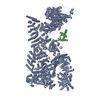 7oikC M: map data used to model this data C: citing same article ( |
|---|---|
| Similar structure data | |
| EM raw data | EMPIAR-10712 (Title: Transmission electron micrographs of mouse RNF213 incubated with ATPγS Data size: 2.3 TB Data #1: unaligned multi-frame micrographs from a dataset collected without grid tilting [micrographs - multiframe] Data #2: unaligned multi-frame micrographs from a dataset collected with a 30 deg grid tilt [micrographs - multiframe]) |

-Links
 PDBj
PDBj


- Assembly
| Deposited unit | 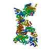
|
|---|---|
| 1 |
|
-Components
-Protein , 1 types, 1 molecules A
| #1: Protein | Mass: 552623.375 Da / Num. of mol.: 1 Source method: isolated from a genetically manipulated source Source: (gene. exp.)  Trichoplusia ni (cabbage looper) Trichoplusia ni (cabbage looper)References: UniProt: E9Q555, RING-type E3 ubiquitin transferase, Hydrolases; Acting on acid anhydrides; Acting on acid anhydrides to facilitate cellular and subcellular movement |
|---|
-Non-polymers , 5 types, 6 molecules 


| #2: Chemical | ChemComp-ATP /  Mass: 507.181 Da / Num. of mol.: 1 / Source method: obtained synthetically / Formula: C10H16N5O13P3 / Feature type: SUBJECT OF INVESTIGATION / Comment: ATP, energy-carrying molecule*YM Mass: 507.181 Da / Num. of mol.: 1 / Source method: obtained synthetically / Formula: C10H16N5O13P3 / Feature type: SUBJECT OF INVESTIGATION / Comment: ATP, energy-carrying molecule*YM | ||||
|---|---|---|---|---|---|
| #3: Chemical | ChemComp-MG /  Mass: 24.305 Da / Num. of mol.: 1 / Source method: obtained synthetically / Formula: Mg / Feature type: SUBJECT OF INVESTIGATION Mass: 24.305 Da / Num. of mol.: 1 / Source method: obtained synthetically / Formula: Mg / Feature type: SUBJECT OF INVESTIGATION | ||||
| #4: Chemical |  Mass: 65.409 Da / Num. of mol.: 2 / Source method: obtained synthetically / Formula: Zn / Feature type: SUBJECT OF INVESTIGATION Mass: 65.409 Da / Num. of mol.: 2 / Source method: obtained synthetically / Formula: Zn / Feature type: SUBJECT OF INVESTIGATION#5: Chemical | ChemComp-ADP / |  Mass: 427.201 Da / Num. of mol.: 1 / Source method: obtained synthetically / Formula: C10H15N5O10P2 / Feature type: SUBJECT OF INVESTIGATION / Comment: ADP, energy-carrying molecule*YM Mass: 427.201 Da / Num. of mol.: 1 / Source method: obtained synthetically / Formula: C10H15N5O10P2 / Feature type: SUBJECT OF INVESTIGATION / Comment: ADP, energy-carrying molecule*YM#6: Chemical | ChemComp-AGS / |  Mass: 523.247 Da / Num. of mol.: 1 / Source method: obtained synthetically / Formula: C10H16N5O12P3S / Feature type: SUBJECT OF INVESTIGATION / Comment: ATP-gamma-S, energy-carrying molecule analogue*YM Mass: 523.247 Da / Num. of mol.: 1 / Source method: obtained synthetically / Formula: C10H16N5O12P3S / Feature type: SUBJECT OF INVESTIGATION / Comment: ATP-gamma-S, energy-carrying molecule analogue*YM |
-Details
| Has ligand of interest | Y |
|---|---|
| Has protein modification | Y |
-Experimental details
-Experiment
| Experiment | Method: ELECTRON MICROSCOPY |
|---|---|
| EM experiment | Aggregation state: PARTICLE / 3D reconstruction method: single particle reconstruction |
- Sample preparation
| Component | Name: RNF213 incubated with ATPgS / Type: COMPLEX / Entity ID: #1 / Source: MULTIPLE SOURCES | ||||||||||||||||
|---|---|---|---|---|---|---|---|---|---|---|---|---|---|---|---|---|---|
| Molecular weight | Value: 0.58 MDa / Experimental value: NO | ||||||||||||||||
| Source (natural) | Organism: | ||||||||||||||||
| Source (recombinant) | Organism: Trichoplusia ni (cabbage looper) | ||||||||||||||||
| Buffer solution | pH: 7.2 | ||||||||||||||||
| Buffer component |
| ||||||||||||||||
| Specimen | Embedding applied: NO / Shadowing applied: NO / Staining applied: NO / Vitrification applied: YES | ||||||||||||||||
| Specimen support | Grid material: GOLD / Grid mesh size: 200 divisions/in. / Grid type: UltrAuFoil R2/2 | ||||||||||||||||
| Vitrification | Instrument: LEICA PLUNGER / Cryogen name: ETHANE |
- Electron microscopy imaging
| Experimental equipment |  Model: Titan Krios / Image courtesy: FEI Company | |||||||||||||||||||||
|---|---|---|---|---|---|---|---|---|---|---|---|---|---|---|---|---|---|---|---|---|---|---|
| Microscopy | Model: TFS KRIOS | |||||||||||||||||||||
| Electron gun | Electron source:  FIELD EMISSION GUN / Accelerating voltage: 300 kV / Illumination mode: SPOT SCAN FIELD EMISSION GUN / Accelerating voltage: 300 kV / Illumination mode: SPOT SCAN | |||||||||||||||||||||
| Electron lens | Mode: BRIGHT FIELD / Nominal defocus max: -2500 nm / Nominal defocus min: -1500 nm / Cs: 2.7 mm / C2 aperture diameter: 50 µm / Alignment procedure: COMA FREE | |||||||||||||||||||||
| Specimen holder | Cryogen: NITROGEN / Specimen holder model: FEI TITAN KRIOS AUTOGRID HOLDER | |||||||||||||||||||||
| Image recording | Imaging-ID: 1 / Num. of grids imaged: 1
| |||||||||||||||||||||
| Image scans | Width: 3838 / Height: 3710 / Movie frames/image: 40 / Used frames/image: 1-40 |
- Processing
| EM software |
| |||||||||||||||||||||||||||||||||||||||||||||
|---|---|---|---|---|---|---|---|---|---|---|---|---|---|---|---|---|---|---|---|---|---|---|---|---|---|---|---|---|---|---|---|---|---|---|---|---|---|---|---|---|---|---|---|---|---|---|
| CTF correction | Type: PHASE FLIPPING AND AMPLITUDE CORRECTION | |||||||||||||||||||||||||||||||||||||||||||||
| Symmetry | Point symmetry: C1 (asymmetric) | |||||||||||||||||||||||||||||||||||||||||||||
| 3D reconstruction | Resolution: 4 Å / Resolution method: FSC 0.143 CUT-OFF / Num. of particles: 336000 / Algorithm: FOURIER SPACE / Num. of class averages: 1 / Symmetry type: POINT | |||||||||||||||||||||||||||||||||||||||||||||
| Atomic model building | Protocol: RIGID BODY FIT / Space: REAL | |||||||||||||||||||||||||||||||||||||||||||||
| Atomic model building | PDB-ID: 6TAX Pdb chain-ID: A / Accession code: 6TAX / Source name: PDB / Type: experimental model |