[English] 日本語
Yorodumi
- PDB-7ofw: Nontypeable Haemophillus influenzae SapA in complex with heme -

+
Open data


ID or keywords:

Loading...

-
Basic information

Entry
Database: PDB / ID: 7ofw
TitleNontypeable Haemophillus influenzae SapA in complex with heme
Components
  • ABC-type transport system, periplasmic component, involved in antimicrobial peptide resistance
  • RNA (5'-R(P*CP*CP*CP*CP*CP*CP*CP*CP*CP*GP*GP*GP*GP*GP*GP*GP*GP*G)-3')
Keywords
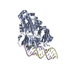
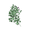
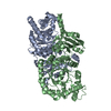
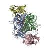
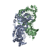
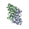
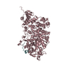
PEPTIDE BINDING PROTEIN / NTHi / RNA binding / peptide binding / substrate binding protein / heme
Function / homology
Function and homology information


ATP-binding cassette (ABC) transporter complex / transmembrane transport / outer membrane-bounded periplasmic space
Similarity search - Function
Solute-binding protein family 5, conserved site / Bacterial extracellular solute-binding proteins, family 5 signature. / Peptide/nickel binding protein, MppA-type / Solute-binding protein family 5 domain / Solute-binding protein family 5 / Bacterial extracellular solute-binding proteins, family 5 Middle
Similarity search - Domain/homology
PROTOPORPHYRIN IX CONTAINING FE / RNA / RNA (> 10) / ABC-type transport system, periplasmic component, involved in antimicrobial peptide resistance
Similarity search - Component
Biological speciesHaemophilus influenzae (bacteria)
Escherichia coli (E. coli)
MethodX-RAY DIFFRACTION / SYNCHROTRON / MOLECULAR REPLACEMENT / molecular replacement / Resolution: 3.15 Å
AuthorsLukacik, P. / Owen, C.D. / Nettleship, J.E. / Bird, L.E. / Owens, R.J. / Walsh, M.A.
Funding support United Kingdom, 1items
OrganizationGrant numberCountry
Medical Research Council (MRC, United Kingdom)MR/M010937/1 United Kingdom
CitationJournal: Plos One / Year: 2021
Title: The structure of nontypeable Haemophilus influenzae SapA in a closed conformation reveals a constricted ligand-binding cavity and a novel RNA binding motif.
Authors: Lukacik, P. / Owen, C.D. / Harris, G. / Bolla, J.R. / Picaud, S. / Alibay, I. / Nettleship, J.E. / Bird, L.E. / Owens, R.J. / Biggin, P.C. / Filippakopoulos, P. / Robinson, C.V. / Walsh, M.A.
History
DepositionMay 5, 2021Deposition site: PDBE / Processing site: PDBE
Revision 1.0Oct 27, 2021Provider: repository / Type: Initial release
Revision 1.1Jan 31, 2024Group: Data collection / Refinement description
Category: chem_comp_atom / chem_comp_bond ...chem_comp_atom / chem_comp_bond / diffrn_source / pdbx_initial_refinement_model
Item: _diffrn_source.pdbx_synchrotron_site

-
Structure visualization

Structure viewerMolecule:
MolmilJmol/JSmol

Downloads & links

-
Assembly

Deposited unit
A: ABC-type transport system, periplasmic component, involved in antimicrobial peptide resistance
C: RNA (5'-R(P*CP*CP*CP*CP*CP*CP*CP*CP*CP*GP*GP*GP*GP*GP*GP*GP*GP*G)-3')
hetero molecules


Theoretical massNumber of molelcules
Total (without water)66,5543
Polymers65,9382
Non-polymers6161
Water362
1
A: ABC-type transport system, periplasmic component, involved in antimicrobial peptide resistance
C: RNA (5'-R(P*CP*CP*CP*CP*CP*CP*CP*CP*CP*GP*GP*GP*GP*GP*GP*GP*GP*G)-3')
hetero molecules

C: RNA (5'-R(P*CP*CP*CP*CP*CP*CP*CP*CP*CP*GP*GP*GP*GP*GP*GP*GP*GP*G)-3')


Theoretical massNumber of molelcules
Total (without water)72,3634
Polymers71,7463
Non-polymers6161
Water181
TypeNameSymmetry operationNumber
identity operation1_555x,y,z1
crystal symmetry operation7_555y,x,-z1
Unit cell
Length a, b, c (Å)142.730, 142.730, 60.580
Angle α, β, γ (deg.)90.000, 90.000, 90.000
Int Tables number90
Space group name H-MP4212
Components on special symmetry positions
IDModelComponents
11A-601-

HEM

21A-601-

HEM

31A-702-

HOH

-
Components

#1: Protein ABC-type transport system, periplasmic component, involved in antimicrobial peptide resistance


Mass: 60129.066 Da / Num. of mol.: 1
Source method: isolated from a genetically manipulated source
Source: (gene. exp.) Haemophilus influenzae (strain 86-028NP) (bacteria)
Strain: 86-028NP / Gene: sapA, NTHI1401 / Plasmid: pOPINF / Production host: Escherichia coli (E. coli) / References: UniProt: Q4QL73
#2: RNA chain RNA (5'-R(P*CP*CP*CP*CP*CP*CP*CP*CP*CP*GP*GP*GP*GP*GP*GP*GP*GP*G)-3')


Mass: 5808.532 Da / Num. of mol.: 1 / Source method: isolated from a natural source / Source: (natural) Escherichia coli (E. coli) / Plasmid details: RNA copurified with the protein
#3: Chemical ChemComp-HEM / PROTOPORPHYRIN IX CONTAINING FE / HEME / Heme B


Mass: 616.487 Da / Num. of mol.: 1 / Source method: obtained synthetically / Formula: C34H32FeN4O4 / Feature type: SUBJECT OF INVESTIGATION
#4: Water ChemComp-HOH / water / Water


Mass: 18.015 Da / Num. of mol.: 2 / Source method: isolated from a natural source / Formula: H2O
Has ligand of interestY

-
Experimental details

-
Experiment

ExperimentMethod: X-RAY DIFFRACTION / Number of used crystals: 1

-
Sample preparation

CrystalDensity Matthews: 2.34 Å3/Da / Density % sol: 47.43 %
Crystal growTemperature: 293 K / Method: vapor diffusion, sitting drop
Details: 22% (w/v) PEG 3350, 0.25 M NaBr, 0.1 M Bis-Tris Propane pH 7.5

-
Data collection

DiffractionMean temperature: 100 K / Serial crystal experiment: N
Diffraction sourceSource: SYNCHROTRON / Site: Diamond / Beamline: I04 / Wavelength: 0.97625 Å
DetectorType: DECTRIS PILATUS 6M-F / Detector: PIXEL / Date: Sep 23, 2014
RadiationProtocol: SINGLE WAVELENGTH / Monochromatic (M) / Laue (L): M / Scattering type: x-ray
Radiation wavelengthWavelength: 0.97625 Å / Relative weight: 1
ReflectionResolution: 3.15→71.47 Å / Num. obs: 11352 / % possible obs: 99.9 % / Redundancy: 5.617 % / Biso Wilson estimate: 79.761 Å2 / CC1/2: 0.997 / Rmerge(I) obs: 0.122 / Rrim(I) all: 0.135 / Χ2: 0.989 / Net I/σ(I): 16.9 / Num. measured all: 116432
Reflection shell

Diffraction-ID: 1

Resolution (Å)Redundancy (%)Rmerge(I) obsMean I/σ(I) obsNum. unique obsCC1/2Rrim(I) all% possible all
3.15-3.235.5990.9792.3815650.7621.08299.9
3.23-3.325.4510.8353.0114820.8390.92599.9
3.32-3.425.7440.6424.1214440.8930.70799.7
3.42-3.525.6250.5165.0314170.9290.5799.7
3.52-3.645.4570.4127.0213470.9420.457100
3.64-3.775.7010.3528.6813330.9460.387100
3.77-3.915.9340.26710.5512600.9740.29399.8
3.91-4.075.8650.20712.4512620.9850.228100
4.07-4.255.5040.15414.3911420.9880.17100
4.25-4.455.8440.1231711440.9920.13699.9
4.45-4.75.7630.09519.6610780.9940.104100
4.7-4.985.3810.08919.99900.9950.09999.9
4.98-5.325.7970.08720.619650.9950.096100
5.32-5.755.6780.08820.128780.9950.097100
5.75-6.35.3780.079208150.9950.088100
6.3-7.045.5790.06523.467370.9960.072100
7.04-8.135.3280.05126.476560.9970.05799.4
8.13-9.965.4560.04131.175460.9980.045100
9.96-14.095.290.0432.494270.9980.044100
14.09-71.474.8920.04232.182410.9980.04799.2

-
Phasing

PhasingMethod: molecular replacement
Phasing MRModel details: Phaser MODE: MR_AUTO
Highest resolutionLowest resolution
Rotation3.15 Å71.36 Å
Translation3.15 Å71.36 Å

-
Processing

Software
NameVersionClassification
XDSdata reduction
XSCALEdata scaling
PHASER2.5.2phasing
REFMAC7.04.09refinement
PDB_EXTRACT3.24data extraction
xia2data reduction
Cootmodel building
BUCCANEERmodel building
BUSTERrefinement
RefinementMethod to determine structure: MOLECULAR REPLACEMENT
Starting model: 3M8U
Resolution: 3.15→71.47 Å / Cor.coef. Fo:Fc: 0.918 / Cor.coef. Fo:Fc free: 0.887 / WRfactor Rfree: 0.2699 / WRfactor Rwork: 0.23 / FOM work R set: 0.803 / SU B: 27.514 / SU ML: 0.451 / SU Rfree: 0.5762 / Cross valid method: THROUGHOUT / σ(F): 0 / ESU R Free: 0.576 / Stereochemistry target values: MAXIMUM LIKELIHOOD
Details: HYDROGENS HAVE BEEN ADDED IN THE RIDING POSITIONS U VALUES : REFINED INDIVIDUALLY
RfactorNum. reflection% reflectionSelection details
Rfree0.2816 553 4.9 %RANDOM
Rwork0.2422 ---
obs0.2441 10777 99.86 %-
Solvent computationIon probe radii: 0.8 Å / Shrinkage radii: 0.8 Å / VDW probe radii: 1.2 Å / Solvent model: MASK
Displacement parametersBiso max: 214.22 Å2 / Biso mean: 93.256 Å2 / Biso min: 39.71 Å2
Baniso -1Baniso -2Baniso -3
1--1 Å20 Å20 Å2
2---1 Å20 Å2
3---2 Å2
Refinement stepCycle: final / Resolution: 3.15→71.47 Å
ProteinNucleic acidLigandSolventTotal
Num. atoms3963 387 43 2 4395
Biso mean--116.03 40.71 -
Num. residues----511
Refine LS restraints
Refine-IDTypeDev idealDev ideal targetNumber
X-RAY DIFFRACTIONr_bond_refined_d0.0070.0144542
X-RAY DIFFRACTIONr_bond_other_d0.0010.0173823
X-RAY DIFFRACTIONr_angle_refined_deg1.3381.6246270
X-RAY DIFFRACTIONr_angle_other_deg0.7521.7078949
X-RAY DIFFRACTIONr_dihedral_angle_1_deg4.6675490
X-RAY DIFFRACTIONr_dihedral_angle_2_deg36.84823.209215
X-RAY DIFFRACTIONr_dihedral_angle_3_deg18.10215683
X-RAY DIFFRACTIONr_dihedral_angle_4_deg21.7631519
X-RAY DIFFRACTIONr_chiral_restr0.0710.2609
X-RAY DIFFRACTIONr_gen_planes_refined0.0050.024819
X-RAY DIFFRACTIONr_gen_planes_other0.0030.02906
LS refinement shellResolution: 3.15→3.232 Å / Rfactor Rfree error: 0 / Total num. of bins used: 20
RfactorNum. reflection% reflection
Rfree0.419 43 -
Rwork0.356 790 -
all-833 -
obs--99.76 %

+
About Yorodumi

-
News

-
Feb 9, 2022. New format data for meta-information of EMDB entries

New format data for meta-information of EMDB entries

  • Version 3 of the EMDB header file is now the official format.
  • The previous official version 1.9 will be removed from the archive.

Related info.:EMDB header

External links:wwPDB to switch to version 3 of the EMDB data model

-
Aug 12, 2020. Covid-19 info

Covid-19 info

URL: https://pdbj.org/emnavi/covid19.php

New page: Covid-19 featured information page in EM Navigator.

Related info.:Covid-19 info / Mar 5, 2020. Novel coronavirus structure data

+
Mar 5, 2020. Novel coronavirus structure data

Novel coronavirus structure data

Related info.:Yorodumi Speices / Aug 12, 2020. Covid-19 info

External links:COVID-19 featured content - PDBj / Molecule of the Month (242):Coronavirus Proteases

+
Jan 31, 2019. EMDB accession codes are about to change! (news from PDBe EMDB page)

EMDB accession codes are about to change! (news from PDBe EMDB page)

  • The allocation of 4 digits for EMDB accession codes will soon come to an end. Whilst these codes will remain in use, new EMDB accession codes will include an additional digit and will expand incrementally as the available range of codes is exhausted. The current 4-digit format prefixed with “EMD-” (i.e. EMD-XXXX) will advance to a 5-digit format (i.e. EMD-XXXXX), and so on. It is currently estimated that the 4-digit codes will be depleted around Spring 2019, at which point the 5-digit format will come into force.
  • The EM Navigator/Yorodumi systems omit the EMD- prefix.

Related info.:Q: What is EMD? / ID/Accession-code notation in Yorodumi/EM Navigator

External links:EMDB Accession Codes are Changing Soon! / Contact to PDBj

+
Jul 12, 2017. Major update of PDB

Major update of PDB

  • wwPDB released updated PDB data conforming to the new PDBx/mmCIF dictionary.
  • This is a major update changing the version number from 4 to 5, and with Remediation, in which all the entries are updated.
  • In this update, many items about electron microscopy experimental information are reorganized (e.g. em_software).
  • Now, EM Navigator and Yorodumi are based on the updated data.

External links:wwPDB Remediation / Enriched Model Files Conforming to OneDep Data Standards Now Available in the PDB FTP Archive

-
Yorodumi

Thousand views of thousand structures

  • Yorodumi is a browser for structure data from EMDB, PDB, SASBDB, etc.
  • This page is also the successor to EM Navigator detail page, and also detail information page/front-end page for Omokage search.
  • The word "yorodu" (or yorozu) is an old Japanese word meaning "ten thousand". "mi" (miru) is to see.

Related info.:EMDB / PDB / SASBDB / Comparison of 3 databanks / Yorodumi Search / Aug 31, 2016. New EM Navigator & Yorodumi / Yorodumi Papers / Jmol/JSmol / Function and homology information / Changes in new EM Navigator and Yorodumi

Read more