[English] 日本語
Yorodumi
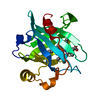
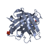
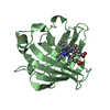
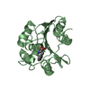
- PDB-7oex: Crystal structure of RBBP9 in complex with phenylalanine -

+
Open data


ID or keywords:

Loading...

-
Basic information

Entry
Database: PDB / ID: 7oex
TitleCrystal structure of RBBP9 in complex with phenylalanine
ComponentsSerine hydrolase RBBP9
KeywordsHYDROLASE / Retinoblastoma-binding protein 9
Function / homology
Function and homology information


type II pneumocyte differentiation / response to nematode / Hydrolases / hydrolase activity / positive regulation of gene expression / nucleoplasm
Similarity search - Function
Putative hydrolase RBBP9/YdeN / Serine hydrolase / Alpha/Beta hydrolase fold
Similarity search - Domain/homology
PHENYLALANINE / Serine hydrolase RBBP9
Similarity search - Component
Biological speciesHomo sapiens (human)
MethodX-RAY DIFFRACTION / SYNCHROTRON / MOLECULAR REPLACEMENT / Resolution: 1.51 Å
AuthorsFiedler, M. / Tang, S. / Chin, J.W.
Funding support United Kingdom, 2items
OrganizationGrant numberCountry
Medical Research Council (MRC, United Kingdom)MC_U105181009 United Kingdom
Medical Research Council (MRC, United Kingdom)MC_UP_A024_1008 United Kingdom
CitationJournal: Nature / Year: 2022
Title: Mechanism-based traps enable protease and hydrolase substrate discovery.
Authors: Tang, S. / Beattie, A.T. / Kafkova, L. / Petris, G. / Huguenin-Dezot, N. / Fiedler, M. / Freeman, M. / Chin, J.W.
History
DepositionMay 4, 2021Deposition site: PDBE / Processing site: PDBE
Revision 1.0Dec 22, 2021Provider: repository / Type: Initial release
Revision 1.1Feb 23, 2022Group: Database references / Category: citation / citation_author
Item: _citation.country / _citation.journal_abbrev ..._citation.country / _citation.journal_abbrev / _citation.journal_id_ASTM / _citation.journal_id_CSD / _citation.journal_id_ISSN / _citation.pdbx_database_id_DOI / _citation.title / _citation.year
Revision 1.2Mar 2, 2022Group: Database references / Category: citation / citation_author
Item: _citation.pdbx_database_id_PubMed / _citation.title ..._citation.pdbx_database_id_PubMed / _citation.title / _citation_author.identifier_ORCID / _citation_author.name
Revision 1.3Mar 9, 2022Group: Database references / Category: citation
Item: _citation.journal_volume / _citation.page_first / _citation.page_last
Revision 1.4Jan 31, 2024Group: Data collection / Refinement description
Category: chem_comp_atom / chem_comp_bond / pdbx_initial_refinement_model

-
Structure visualization

Structure viewerMolecule:
MolmilJmol/JSmol

Downloads & links

-
Assembly

Deposited unit
A: Serine hydrolase RBBP9
B: Serine hydrolase RBBP9
hetero molecules


Theoretical massNumber of molelcules
Total (without water)44,5244
Polymers44,1942
Non-polymers3302
Water5,260292
1
A: Serine hydrolase RBBP9
hetero molecules


Theoretical massNumber of molelcules
Total (without water)22,2622
Polymers22,0971
Non-polymers1651
Water181
TypeNameSymmetry operationNumber
identity operation1_555x,y,z1
2
B: Serine hydrolase RBBP9
hetero molecules


Theoretical massNumber of molelcules
Total (without water)22,2622
Polymers22,0971
Non-polymers1651
Water181
TypeNameSymmetry operationNumber
identity operation1_555x,y,z1
Unit cell
Length a, b, c (Å)36.956, 130.127, 38.993
Angle α, β, γ (deg.)90.000, 115.240, 90.000
Int Tables number4
Space group name H-MP1211

-
Components

#1: Protein Serine hydrolase RBBP9 / B5T-overexpressed gene protein / Protein BOG / Retinoblastoma-binding protein 10 / RBBP-10 / ...B5T-overexpressed gene protein / Protein BOG / Retinoblastoma-binding protein 10 / RBBP-10 / Retinoblastoma-binding protein 9 / RBBP-9


Mass: 22097.057 Da / Num. of mol.: 2
Source method: isolated from a genetically manipulated source
Source: (gene. exp.) Homo sapiens (human) / Gene: RBBP9, BOG, RBBP10 / Production host: Escherichia coli BL21(DE3) (bacteria) / References: UniProt: O75884, Hydrolases
#2: Chemical ChemComp-PHE / PHENYLALANINE


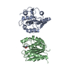
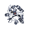
Type: L-peptide linking / Mass: 165.189 Da / Num. of mol.: 2 / Source method: isolated from a natural source / Formula: C9H11NO2 / Feature type: SUBJECT OF INVESTIGATION
#3: Water ChemComp-HOH / water


Mass: 18.015 Da / Num. of mol.: 292 / Source method: isolated from a natural source / Formula: H2O
Has ligand of interestY

-
Experimental details

-
Experiment

ExperimentMethod: X-RAY DIFFRACTION / Number of used crystals: 1

-
Sample preparation

CrystalDensity Matthews: 1.92 Å3/Da / Density % sol: 35.9 %
Crystal growTemperature: 291 K / Method: vapor diffusion, sitting drop / pH: 6.5 / Details: 30% w/v PEG 4K, 0.1 M MES sodium salt pH 6.5

-
Data collection

DiffractionMean temperature: 130 K / Serial crystal experiment: N
Diffraction sourceSource: SYNCHROTRON / Site: Diamond / Beamline: I04 / Wavelength: 0.9795 Å
DetectorType: DECTRIS EIGER2 XE 16M / Detector: PIXEL / Date: Feb 3, 2020
RadiationProtocol: SINGLE WAVELENGTH / Monochromatic (M) / Laue (L): M / Scattering type: x-ray
Radiation wavelengthWavelength: 0.9795 Å / Relative weight: 1
ReflectionResolution: 1.51→29.75 Å / Num. obs: 49673 / % possible obs: 96.1 % / Redundancy: 7 % / CC1/2: 0.999 / Rmerge(I) obs: 0.068 / Rpim(I) all: 0.028 / Rrim(I) all: 0.073 / Net I/σ(I): 14
Reflection shell

Diffraction-ID: 1

Resolution (Å)Redundancy (%)Rmerge(I) obsNum. measured allNum. unique obsCC1/2Rpim(I) allRrim(I) allNet I/σ(I) obs% possible all
1.51-1.545.10.669939218410.8130.3120.7422.372
8.29-29.7370.03422323200.9990.0130.03642.296.3

-
Processing

Software
NameVersionClassification
Aimless0.7.4data scaling
REFMAC5.8.0267refinement
PDB_EXTRACT3.27data extraction
xia2data reduction
PHASERphasing
RefinementMethod to determine structure: MOLECULAR REPLACEMENT
Starting model: 2QS9
Resolution: 1.51→29.73 Å / Cor.coef. Fo:Fc: 0.972 / Cor.coef. Fo:Fc free: 0.958 / SU B: 1.688 / SU ML: 0.059 / SU R Cruickshank DPI: 0.0785 / Cross valid method: THROUGHOUT / ESU R: 0.079 / ESU R Free: 0.082 / Stereochemistry target values: MAXIMUM LIKELIHOOD
Details: HYDROGENS HAVE BEEN ADDED IN THE RIDING POSITIONS U VALUES : REFINED INDIVIDUALLY
RfactorNum. reflection% reflectionSelection details
Rfree0.1972 2383 4.8 %RANDOM
Rwork0.161 ---
obs0.1627 47250 95.93 %-
Solvent computationIon probe radii: 0.7 Å / Shrinkage radii: 0.7 Å / VDW probe radii: 1.1 Å / Solvent model: MASK
Displacement parametersBiso max: 134.29 Å2 / Biso mean: 20.106 Å2 / Biso min: 7.07 Å2
Baniso -1Baniso -2Baniso -3
1--0.08 Å20 Å20.08 Å2
2--0.02 Å2-0 Å2
3----0.01 Å2
Refinement stepCycle: final / Resolution: 1.51→29.73 Å
ProteinNucleic acidLigandSolventTotal
Num. atoms3006 0 24 292 3322
Biso mean--21.77 29.06 -
Num. residues----376
Refine LS restraints
Refine-IDTypeDev idealDev ideal targetNumber
X-RAY DIFFRACTIONr_bond_refined_d0.0110.0183121
X-RAY DIFFRACTIONr_bond_other_d0.0010.0192852
X-RAY DIFFRACTIONr_angle_refined_deg1.4331.8424241
X-RAY DIFFRACTIONr_angle_other_deg1.1632.7356605
X-RAY DIFFRACTIONr_dihedral_angle_1_deg5.9235374
X-RAY DIFFRACTIONr_dihedral_angle_2_deg35.25723.013156
X-RAY DIFFRACTIONr_dihedral_angle_3_deg10.44215509
X-RAY DIFFRACTIONr_dihedral_angle_4_deg15.3631512
X-RAY DIFFRACTIONr_chiral_restr0.0940.2451
X-RAY DIFFRACTIONr_gen_planes_refined0.0070.023492
X-RAY DIFFRACTIONr_gen_planes_other0.0010.02704
X-RAY DIFFRACTIONr_nbd_refined
X-RAY DIFFRACTIONr_nbd_other
X-RAY DIFFRACTIONr_nbtor_refined
X-RAY DIFFRACTIONr_nbtor_other
X-RAY DIFFRACTIONr_xyhbond_nbd_refined
X-RAY DIFFRACTIONr_xyhbond_nbd_other
X-RAY DIFFRACTIONr_metal_ion_refined
X-RAY DIFFRACTIONr_metal_ion_other
X-RAY DIFFRACTIONr_symmetry_vdw_refined
X-RAY DIFFRACTIONr_symmetry_vdw_other
X-RAY DIFFRACTIONr_symmetry_hbond_refined
X-RAY DIFFRACTIONr_symmetry_hbond_other
X-RAY DIFFRACTIONr_symmetry_metal_ion_refined
X-RAY DIFFRACTIONr_symmetry_metal_ion_other
X-RAY DIFFRACTIONr_rigid_bond_restr
X-RAY DIFFRACTIONr_sphericity_free
X-RAY DIFFRACTIONr_sphericity_bonded
LS refinement shellResolution: 1.513→1.552 Å / Rfactor Rfree error: 0
RfactorNum. reflection% reflection
Rfree0.317 154 -
Rwork0.317 2829 -
obs--78.03 %

+
About Yorodumi

-
News

-
Feb 9, 2022. New format data for meta-information of EMDB entries

New format data for meta-information of EMDB entries

  • Version 3 of the EMDB header file is now the official format.
  • The previous official version 1.9 will be removed from the archive.

Related info.:EMDB header

External links:wwPDB to switch to version 3 of the EMDB data model

-
Aug 12, 2020. Covid-19 info

Covid-19 info

URL: https://pdbjlvh1.pdbj.org/emnavi/covid19.php

New page: Covid-19 featured information page in EM Navigator.

Related info.:Covid-19 info / Mar 5, 2020. Novel coronavirus structure data

+
Mar 5, 2020. Novel coronavirus structure data

Novel coronavirus structure data

Related info.:Yorodumi Speices / Aug 12, 2020. Covid-19 info

External links:COVID-19 featured content - PDBj / Molecule of the Month (242):Coronavirus Proteases

+
Jan 31, 2019. EMDB accession codes are about to change! (news from PDBe EMDB page)

EMDB accession codes are about to change! (news from PDBe EMDB page)

  • The allocation of 4 digits for EMDB accession codes will soon come to an end. Whilst these codes will remain in use, new EMDB accession codes will include an additional digit and will expand incrementally as the available range of codes is exhausted. The current 4-digit format prefixed with “EMD-” (i.e. EMD-XXXX) will advance to a 5-digit format (i.e. EMD-XXXXX), and so on. It is currently estimated that the 4-digit codes will be depleted around Spring 2019, at which point the 5-digit format will come into force.
  • The EM Navigator/Yorodumi systems omit the EMD- prefix.

Related info.:Q: What is EMD? / ID/Accession-code notation in Yorodumi/EM Navigator

External links:EMDB Accession Codes are Changing Soon! / Contact to PDBj

+
Jul 12, 2017. Major update of PDB

Major update of PDB

  • wwPDB released updated PDB data conforming to the new PDBx/mmCIF dictionary.
  • This is a major update changing the version number from 4 to 5, and with Remediation, in which all the entries are updated.
  • In this update, many items about electron microscopy experimental information are reorganized (e.g. em_software).
  • Now, EM Navigator and Yorodumi are based on the updated data.

External links:wwPDB Remediation / Enriched Model Files Conforming to OneDep Data Standards Now Available in the PDB FTP Archive

-
Yorodumi

Thousand views of thousand structures

  • Yorodumi is a browser for structure data from EMDB, PDB, SASBDB, etc.
  • This page is also the successor to EM Navigator detail page, and also detail information page/front-end page for Omokage search.
  • The word "yorodu" (or yorozu) is an old Japanese word meaning "ten thousand". "mi" (miru) is to see.

Related info.:EMDB / PDB / SASBDB / Comparison of 3 databanks / Yorodumi Search / Aug 31, 2016. New EM Navigator & Yorodumi / Yorodumi Papers / Jmol/JSmol / Function and homology information / Changes in new EM Navigator and Yorodumi

Read more