 Movie
Movie Controller

[English] 日本語
 Yorodumi
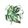
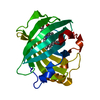
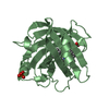
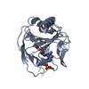
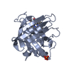
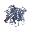
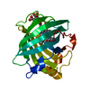
- PDB-3np1: CRYSTAL STRUCTURE OF THE COMPLEX OF NITROPHORIN 1 FROM RHODNIUS P... -
+ Open data

- Basic information
| Entry | Database: PDB / ID: 3np1 | ||||||
|---|---|---|---|---|---|---|---|
| Title | CRYSTAL STRUCTURE OF THE COMPLEX OF NITROPHORIN 1 FROM RHODNIUS PROLIXUS WITH CYANIDE | ||||||
 Components Components | NITROPHORIN 1 | ||||||
 Keywords Keywords | NITRIC OXIDE TRANSPORT / FERRIC HEME / HISTAMINE / ANTIHISTAMINE / VASODILATOR / LIPOCALIN / CYANIDE | ||||||
| Function / homology |  Function and homology information Function and homology informationhistamine binding / nitric oxide binding / vasodilation / extracellular region / metal ion binding Similarity search - Function | ||||||
| Biological species |  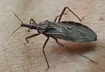 Rhodnius prolixus (insect) Rhodnius prolixus (insect) | ||||||
| Method |  X-RAY DIFFRACTION / DIFFERENCE FOURIER / Resolution: 2.3 Å X-RAY DIFFRACTION / DIFFERENCE FOURIER / Resolution: 2.3 Å | ||||||
 Authors Authors | Weichsel, A. / Andersen, J.F. / Champagne, D.E. / Walker, F.A. / Montfort, W.R. | ||||||
 Citation Citation | Journal: Nat.Struct.Biol. / Year: 1998 Title: Crystal structures of a nitric oxide transport protein from a blood-sucking insect. Authors: Weichsel, A. / Andersen, J.F. / Champagne, D.E. / Walker, F.A. / Montfort, W.R. #1: Journal: Biochemistry / Year: 1997Title: Nitric Oxide Binding and Crystallization of Recombinant Nitrophorin I, a Nitric Oxide Transport Protein from the Blood-Sucking Bug Rhodnius Prolixus Authors: Andersen, J.F. / Champagne, D.E. / Weichsel, A. / Ribeiro, J.M. / Balfour, C.A. / Dress, V. / Montfort, W.R. | ||||||
| History |
|
- Structure visualization
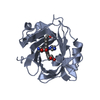
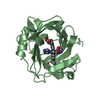
| Structure viewer | Molecule: MolmilJmol/JSmol |
|---|
- Downloads & links
-Download
| PDBx/mmCIF format | 3np1.cif.gz | 88.8 KB | Display | PDBx/mmCIF format |
|---|---|---|---|---|
| PDB format | pdb3np1.ent.gz | 68.1 KB | Display | PDB format |
| PDBx/mmJSON format | 3np1.json.gz | Tree view | PDBx/mmJSON format | |
| Others |  Other downloads Other downloads |
-Validation report
| Arichive directory | https://data.pdbj.org/pub/pdb/validation_reports/np/3np1ftp://data.pdbj.org/pub/pdb/validation_reports/np/3np1 | HTTPS FTP |
|---|
-Related structure data
| Related structure data |  1np1SC 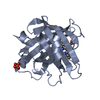 2np1C S: Starting model for refinement C: citing same article ( |
|---|---|
| Similar structure data |

-Links
 PDBj
PDBj


- Assembly
| Deposited unit | 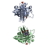
| ||||||||
|---|---|---|---|---|---|---|---|---|---|
| 1 | 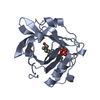
| ||||||||
| 2 | 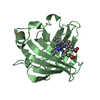
| ||||||||
| Unit cell |
| ||||||||
| Noncrystallographic symmetry (NCS) | NCS oper: (Code: given Matrix: (0.697379, -0.038635, -0.715661), Vector: |
-Components
| #1: Protein | Mass: 20510.867 Da / Num. of mol.: 2 Source method: isolated from a genetically manipulated source Source: (gene. exp.) Rhodnius prolixus (insect) / Cell line: BL21 / Organ: SALIVARY GLAND / Plasmid: PET17B-NP1 / Species (production host): Escherichia coli / Production host:  #2: Chemical |   Mass: 26.017 Da / Num. of mol.: 2 / Source method: obtained synthetically / Formula: CN Mass: 26.017 Da / Num. of mol.: 2 / Source method: obtained synthetically / Formula: CN#3: Chemical |   Mass: 94.971 Da / Num. of mol.: 2 / Source method: obtained synthetically / Formula: PO4 Mass: 94.971 Da / Num. of mol.: 2 / Source method: obtained synthetically / Formula: PO4#4: Chemical |   Mass: 616.487 Da / Num. of mol.: 2 / Source method: obtained synthetically / Formula: C34H32FeN4O4 Mass: 616.487 Da / Num. of mol.: 2 / Source method: obtained synthetically / Formula: C34H32FeN4O4#5: Water | ChemComp-HOH / |  Mass: 18.015 Da / Num. of mol.: 120 / Source method: isolated from a natural source / Formula: H2O Mass: 18.015 Da / Num. of mol.: 120 / Source method: isolated from a natural source / Formula: H2OHas protein modification | Y | |
|---|
-Experimental details
-Experiment
| Experiment | Method: X-RAY DIFFRACTION / Number of used crystals: 1 |
|---|
- Sample preparation
| Crystal | Density Matthews: 2.4 Å3/Da / Density % sol: 48 % | |||||||||||||||||||||||||
|---|---|---|---|---|---|---|---|---|---|---|---|---|---|---|---|---|---|---|---|---|---|---|---|---|---|---|
| Crystal grow | pH: 7.5 Details: 2.8 M AMMONIUM PHOSPHATE, 0.1 M TRIS.HCL, PH 7.5, ROOM TEMPERATURE | |||||||||||||||||||||||||
| Crystal grow | *PLUS Method: vapor diffusion, hanging drop / Details: Andersen, J.F., (1997) Biochemistry, 36, 4423. | |||||||||||||||||||||||||
| Components of the solutions | *PLUS
|
-Data collection
| Diffraction | Mean temperature: 293 K |
|---|---|
| Diffraction source | Source: ROTATING ANODE / Type: ENRAF-NONIUS FR571 / Wavelength: 1.5418 |
| Detector | Type: ENRAF-NONIUS FAST / Detector: DIFFRACTOMETER / Date: Oct 17, 1997 / Details: COLLIMATOR |
| Radiation | Monochromator: GRAPHITE(002) / Monochromatic (M) / Laue (L): M / Scattering type: x-ray |
| Radiation wavelength | Wavelength: 1.5418 Å / Relative weight: 1 |
| Reflection | Resolution: 2.3→28 Å / Num. obs: 16620 / % possible obs: 99 % / Observed criterion σ(I): 0 / Redundancy: 2 % / Biso Wilson estimate: 25 Å2 / Rsym value: 0.056 / Net I/σ(I): 10 |
| Reflection shell | Resolution: 2.3→2.4 Å / Redundancy: 1.9 % / Mean I/σ(I) obs: 2.8 / Rsym value: 0.22 / % possible all: 97.2 |
| Reflection | *PLUS Num. measured all: 46715 / Rmerge(I) obs: 0.053 |
- Processing
| Software |
| ||||||||||||||||||||||||||||||||||||||||||||||||||||||||||||||||||||||||||||||||
|---|---|---|---|---|---|---|---|---|---|---|---|---|---|---|---|---|---|---|---|---|---|---|---|---|---|---|---|---|---|---|---|---|---|---|---|---|---|---|---|---|---|---|---|---|---|---|---|---|---|---|---|---|---|---|---|---|---|---|---|---|---|---|---|---|---|---|---|---|---|---|---|---|---|---|---|---|---|---|---|---|---|
| Refinement | Method to determine structure: DIFFERENCE FOURIER Starting model: PDB ENTRY 1NP1 Resolution: 2.3→15 Å / Rfactor Rfree error: 0.013 / Data cutoff low absF: 0.1 / Isotropic thermal model: RESTRAINED / Cross valid method: THROUGHOUT / σ(F): 0 / Details: BULK SOLVENT CORRECTION WITH K = 0.3435, B = 94.9.
| ||||||||||||||||||||||||||||||||||||||||||||||||||||||||||||||||||||||||||||||||
| Displacement parameters | Biso mean: 32 Å2 | ||||||||||||||||||||||||||||||||||||||||||||||||||||||||||||||||||||||||||||||||
| Refine analyze | Luzzati sigma a obs: 0.42 Å | ||||||||||||||||||||||||||||||||||||||||||||||||||||||||||||||||||||||||||||||||
| Refinement step | Cycle: LAST / Resolution: 2.3→15 Å
| ||||||||||||||||||||||||||||||||||||||||||||||||||||||||||||||||||||||||||||||||
| Refine LS restraints |
| ||||||||||||||||||||||||||||||||||||||||||||||||||||||||||||||||||||||||||||||||
| Refine LS restraints NCS | NCS model details: UNRESTRAINED | ||||||||||||||||||||||||||||||||||||||||||||||||||||||||||||||||||||||||||||||||
| LS refinement shell | Resolution: 2.3→2.4 Å / Rfactor Rfree error: 0.081 / Total num. of bins used: 8
| ||||||||||||||||||||||||||||||||||||||||||||||||||||||||||||||||||||||||||||||||
| Xplor file |
| ||||||||||||||||||||||||||||||||||||||||||||||||||||||||||||||||||||||||||||||||
| Software | *PLUS Name: X-PLOR / Version: 3.851 / Classification: refinement | ||||||||||||||||||||||||||||||||||||||||||||||||||||||||||||||||||||||||||||||||
| Refine LS restraints | *PLUS
|
