 Movie
Movie Controller

[English] 日本語
 Yorodumi
- PDB-7ocs: Mannitol-1-phosphate bound to the phosphatase domain of the bifun... -
+ Open data

- Basic information
| Entry | Database: PDB / ID: 7ocs | ||||||
|---|---|---|---|---|---|---|---|
| Title | Mannitol-1-phosphate bound to the phosphatase domain of the bifunctional mannitol-1-phosphate dehydrogenase/phosphatase MtlD-D16A from Acinetobacter baumannii | ||||||
 Components Components | HAD hydrolase, family IA, variant 3 | ||||||
 Keywords Keywords | OXIDOREDUCTASE / Mannitol / Dehydrogenase / Phosphatase / NADPH / Fructose-6-phosphate / HAD hydrolase family IA variant 3 | ||||||
| Function / homology |  Function and homology information Function and homology informationoxidoreductase activity / nucleotide binding / hydrolase activity / metal ion binding Similarity search - Function | ||||||
| Biological species |  Acinetobacter baumannii ATCC 19606 = CIP 70.34 = JCM 6841 (bacteria) Acinetobacter baumannii ATCC 19606 = CIP 70.34 = JCM 6841 (bacteria) | ||||||
| Method |  X-RAY DIFFRACTION / SYNCHROTRON / MOLECULAR REPLACEMENT / Resolution: 2.3 Å X-RAY DIFFRACTION / SYNCHROTRON / MOLECULAR REPLACEMENT / Resolution: 2.3 Å | ||||||
 Authors Authors | Tam, H.K. / Mueller, V. / Pos, K.M. | ||||||
| Funding support |  Germany, 1items Germany, 1items
| ||||||
 Citation Citation | Journal: Proc.Natl.Acad.Sci.USA / Year: 2022 Title: Unidirectional mannitol synthesis of Acinetobacter baumannii MtlD is facilitated by the helix-loop-helix-mediated dimer formation. Authors: Tam, H.K. / Konig, P. / Himpich, S. / Ngu, N.D. / Abele, R. / Muller, V. / Pos, K.M. | ||||||
| History |
|
- Structure visualization
| Structure viewer | Molecule: MolmilJmol/JSmol |
|---|
- Downloads & links
-Download
| PDBx/mmCIF format | 7ocs.cif.gz | 1 MB | Display | PDBx/mmCIF format |
|---|---|---|---|---|
| PDB format | pdb7ocs.ent.gz | 873.4 KB | Display | PDB format |
| PDBx/mmJSON format | 7ocs.json.gz | Tree view | PDBx/mmJSON format | |
| Others |  Other downloads Other downloads |
-Validation report
| Arichive directory | https://data.pdbj.org/pub/pdb/validation_reports/oc/7ocsftp://data.pdbj.org/pub/pdb/validation_reports/oc/7ocs | HTTPS FTP |
|---|
-Related structure data
| Related structure data | 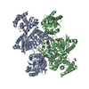 7ocnSC 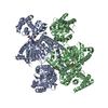 7ocpC 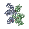 7ocqC 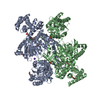 7ocrC 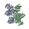 7octC 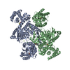 7ocuC S: Starting model for refinement C: citing same article ( |
|---|---|
| Similar structure data |
-Links
 PDBj
PDBj


- Assembly
| Deposited unit | 
| ||||||||
|---|---|---|---|---|---|---|---|---|---|
| 1 | 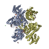
| ||||||||
| 2 | 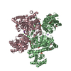
| ||||||||
| Unit cell |
|
-Components
-Protein , 1 types, 4 molecules ABCD
| #1: Protein | Mass: 83504.250 Da / Num. of mol.: 4 Source method: isolated from a genetically manipulated source Source: (gene. exp.) Acinetobacter baumannii ATCC 19606 = CIP 70.34 = JCM 6841 (bacteria)Strain: ATCC 19606 / DSM 30007 / CIP 70.34 / JCM 6841 / NBRC 109757 / NCIMB 12457 / NCTC 12156 / 81 Gene: HMPREF0010_00722 / Plasmid: PLASMID / Details (production host): pET21 / Production host: |
|---|
-Non-polymers , 13 types, 622 molecules 


| #2: Chemical |  Mass: 92.094 Da / Num. of mol.: 2 / Source method: obtained synthetically / Formula: C3H8O3 Mass: 92.094 Da / Num. of mol.: 2 / Source method: obtained synthetically / Formula: C3H8O3#3: Chemical | ChemComp-EDO /  Mass: 62.068 Da / Num. of mol.: 13 / Source method: obtained synthetically / Formula: C2H6O2 Mass: 62.068 Da / Num. of mol.: 13 / Source method: obtained synthetically / Formula: C2H6O2#4: Chemical |  Mass: 150.173 Da / Num. of mol.: 2 / Source method: obtained synthetically / Formula: C6H14O4 Mass: 150.173 Da / Num. of mol.: 2 / Source method: obtained synthetically / Formula: C6H14O4#5: Chemical |  Mass: 194.226 Da / Num. of mol.: 2 / Source method: obtained synthetically / Formula: C8H18O5 / Comment: precipitant*YM Mass: 194.226 Da / Num. of mol.: 2 / Source method: obtained synthetically / Formula: C8H18O5 / Comment: precipitant*YM#6: Chemical |  Mass: 78.133 Da / Num. of mol.: 2 / Source method: obtained synthetically / Formula: C2H6OS Mass: 78.133 Da / Num. of mol.: 2 / Source method: obtained synthetically / Formula: C2H6OS#7: Chemical | ChemComp-SO4 /  Mass: 96.063 Da / Num. of mol.: 10 / Source method: obtained synthetically / Formula: SO4 Mass: 96.063 Da / Num. of mol.: 10 / Source method: obtained synthetically / Formula: SO4#8: Chemical | ChemComp-MG / |  Mass: 24.305 Da / Num. of mol.: 1 / Source method: obtained synthetically / Formula: Mg Mass: 24.305 Da / Num. of mol.: 1 / Source method: obtained synthetically / Formula: Mg#9: Chemical | ChemComp-CL /  Mass: 35.453 Da / Num. of mol.: 10 / Source method: obtained synthetically / Formula: Cl Mass: 35.453 Da / Num. of mol.: 10 / Source method: obtained synthetically / Formula: Cl#10: Chemical | ChemComp-44H /  Mass: 262.152 Da / Num. of mol.: 4 / Source method: obtained synthetically / Formula: C6H15O9P Mass: 262.152 Da / Num. of mol.: 4 / Source method: obtained synthetically / Formula: C6H15O9P#11: Chemical | ChemComp-EPE / |  Mass: 238.305 Da / Num. of mol.: 1 / Source method: obtained synthetically / Formula: C8H18N2O4S / Comment: pH buffer*YM Mass: 238.305 Da / Num. of mol.: 1 / Source method: obtained synthetically / Formula: C8H18N2O4S / Comment: pH buffer*YM#12: Chemical |  Mass: 22.990 Da / Num. of mol.: 2 / Source method: obtained synthetically / Formula: Na Mass: 22.990 Da / Num. of mol.: 2 / Source method: obtained synthetically / Formula: Na#13: Chemical | ChemComp-ACT / |  Mass: 59.044 Da / Num. of mol.: 1 / Source method: obtained synthetically / Formula: C2H3O2 Mass: 59.044 Da / Num. of mol.: 1 / Source method: obtained synthetically / Formula: C2H3O2#14: Water | ChemComp-HOH / | Mass: 18.015 Da / Num. of mol.: 572 / Source method: isolated from a natural source / Formula: H2O |
|---|
-Details
| Has ligand of interest | Y |
|---|
-Experimental details
-Experiment
| Experiment | Method: X-RAY DIFFRACTION / Number of used crystals: 1 |
|---|
- Sample preparation
| Crystal | Density Matthews: 2.62 Å3/Da / Density % sol: 53.16 % |
|---|---|
| Crystal grow | Temperature: 291 K / Method: vapor diffusion, sitting drop / pH: 6.5 Details: .1M BIS-Tris propane pH6.5, 0.15M Na2SO4, 14% PEG3350, 0.02M MgCl2, 0.1M Potassium acetate with microseeding |
-Data collection
| Diffraction | Mean temperature: 100 K / Serial crystal experiment: N | ||||||||||||||||||||||||||||||
|---|---|---|---|---|---|---|---|---|---|---|---|---|---|---|---|---|---|---|---|---|---|---|---|---|---|---|---|---|---|---|---|
| Diffraction source | Source: SYNCHROTRON / Site: SOLEIL  / Beamline: PROXIMA 2 / Wavelength: 0.9788 Å / Beamline: PROXIMA 2 / Wavelength: 0.9788 Å | ||||||||||||||||||||||||||||||
| Detector | Type: DECTRIS EIGER X 9M / Detector: PIXEL / Date: Apr 27, 2019 | ||||||||||||||||||||||||||||||
| Radiation | Protocol: SINGLE WAVELENGTH / Monochromatic (M) / Laue (L): M / Scattering type: x-ray | ||||||||||||||||||||||||||||||
| Radiation wavelength | Wavelength: 0.9788 Å / Relative weight: 1 | ||||||||||||||||||||||||||||||
| Reflection | Resolution: 2.3→48.61 Å / Num. obs: 149396 / % possible obs: 100 % / Redundancy: 7.1 % / Biso Wilson estimate: 41.03 Å2 / CC1/2: 0.993 / Rmerge(I) obs: 0.22 / Rpim(I) all: 0.088 / Rrim(I) all: 0.237 / Net I/σ(I): 5.3 / Num. measured all: 1060197 / Scaling rejects: 661 | ||||||||||||||||||||||||||||||
| Reflection shell | Diffraction-ID: 1
|
- Processing
| Software |
| |||||||||||||||||||||||||||||||||||||||||||||||||||||||||||||||||||||||||||||||||||||||||||||||||||||||||||||||||||||||||||||
|---|---|---|---|---|---|---|---|---|---|---|---|---|---|---|---|---|---|---|---|---|---|---|---|---|---|---|---|---|---|---|---|---|---|---|---|---|---|---|---|---|---|---|---|---|---|---|---|---|---|---|---|---|---|---|---|---|---|---|---|---|---|---|---|---|---|---|---|---|---|---|---|---|---|---|---|---|---|---|---|---|---|---|---|---|---|---|---|---|---|---|---|---|---|---|---|---|---|---|---|---|---|---|---|---|---|---|---|---|---|---|---|---|---|---|---|---|---|---|---|---|---|---|---|---|---|---|
| Refinement | Method to determine structure: MOLECULAR REPLACEMENT Starting model: 7OCN Resolution: 2.3→48.61 Å / Cor.coef. Fo:Fc: 0.889 / Cor.coef. Fo:Fc free: 0.863 / SU R Cruickshank DPI: 0.297 / Cross valid method: THROUGHOUT / σ(F): 0 / SU R Blow DPI: 0.281 / SU Rfree Blow DPI: 0.211 / SU Rfree Cruickshank DPI: 0.218
| |||||||||||||||||||||||||||||||||||||||||||||||||||||||||||||||||||||||||||||||||||||||||||||||||||||||||||||||||||||||||||||
| Displacement parameters | Biso max: 135.44 Å2 / Biso mean: 53.47 Å2 / Biso min: 17.09 Å2
| |||||||||||||||||||||||||||||||||||||||||||||||||||||||||||||||||||||||||||||||||||||||||||||||||||||||||||||||||||||||||||||
| Refine analyze | Luzzati coordinate error obs: 0.37 Å | |||||||||||||||||||||||||||||||||||||||||||||||||||||||||||||||||||||||||||||||||||||||||||||||||||||||||||||||||||||||||||||
| Refinement step | Cycle: final / Resolution: 2.3→48.61 Å
| |||||||||||||||||||||||||||||||||||||||||||||||||||||||||||||||||||||||||||||||||||||||||||||||||||||||||||||||||||||||||||||
| Refine LS restraints |
| |||||||||||||||||||||||||||||||||||||||||||||||||||||||||||||||||||||||||||||||||||||||||||||||||||||||||||||||||||||||||||||
| LS refinement shell | Resolution: 2.3→2.32 Å / Rfactor Rfree error: 0 / Total num. of bins used: 51
| |||||||||||||||||||||||||||||||||||||||||||||||||||||||||||||||||||||||||||||||||||||||||||||||||||||||||||||||||||||||||||||
| Refinement TLS params. | Method: refined / Refine-ID: X-RAY DIFFRACTION
| |||||||||||||||||||||||||||||||||||||||||||||||||||||||||||||||||||||||||||||||||||||||||||||||||||||||||||||||||||||||||||||
| Refinement TLS group |
|
