 Movie
Movie Controller

[English] 日本語
 Yorodumi
- PDB-7o9o: Crystal structure of the Awp3b (adhesin-like wall protein 3b) A-d... -
+ Open data

- Basic information
| Entry | Database: PDB / ID: 7o9o | ||||||
|---|---|---|---|---|---|---|---|
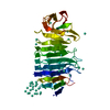
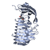
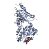
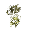
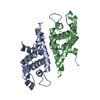
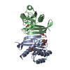
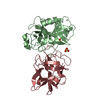
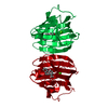
| Title | Crystal structure of the Awp3b (adhesin-like wall protein 3b) A-domain from Candida glabrata | ||||||
 Components Components | AWP3b | ||||||
 Keywords Keywords | CELL ADHESION / Candida glabrata / putative adhesin / adhesin-like wall protein / Awp / adhesion / beta-helix / haze-protective factor / cell wall protein | ||||||
| Function / homology | AWP3b Function and homology information Function and homology information | ||||||
| Biological species |  Candida glabrata (fungus) Candida glabrata (fungus) | ||||||
| Method |  X-RAY DIFFRACTION / SYNCHROTRON / MOLECULAR REPLACEMENT / Resolution: 1.55 Å X-RAY DIFFRACTION / SYNCHROTRON / MOLECULAR REPLACEMENT / Resolution: 1.55 Å | ||||||
 Authors Authors | Reithofer, V. / de Groot, P. / Essen, L.-O. | ||||||
| Funding support |  Germany, 1items Germany, 1items
| ||||||
 Citation Citation | Journal: Plos Pathog. / Year: 2021 Title: A novel class of Candida glabrata cell wall proteins with beta-helix fold mediates adhesion in clinical isolates. Authors: Reithofer, V. / Fernandez-Pereira, J. / Alvarado, M. / de Groot, P. / Essen, L.O. | ||||||
| History |
|
- Structure visualization
| Structure viewer | Molecule: MolmilJmol/JSmol |
|---|
- Downloads & links
-Download
| PDBx/mmCIF format | 7o9o.cif.gz | 247.5 KB | Display | PDBx/mmCIF format |
|---|---|---|---|---|
| PDB format | pdb7o9o.ent.gz | 164.4 KB | Display | PDB format |
| PDBx/mmJSON format | 7o9o.json.gz | Tree view | PDBx/mmJSON format | |
| Others |  Other downloads Other downloads |
-Validation report
| Summary document | 7o9o_validation.pdf.gz | 419.6 KB | Display | wwPDB validaton report |
|---|---|---|---|---|
| Full document | 7o9o_full_validation.pdf.gz | 419.8 KB | Display | |
| Data in XML | 7o9o_validation.xml.gz | 19.4 KB | Display | |
| Data in CIF | 7o9o_validation.cif.gz | 31.4 KB | Display | |
| Arichive directory | https://data.pdbj.org/pub/pdb/validation_reports/o9/7o9oftp://data.pdbj.org/pub/pdb/validation_reports/o9/7o9o | HTTPS FTP |
-Related structure data

-Links
 PDBj
PDBj- Assembly
| Deposited unit | 
| ||||||||||||
|---|---|---|---|---|---|---|---|---|---|---|---|---|---|
| 1 |
| ||||||||||||
| Unit cell |
| ||||||||||||
| Components on special symmetry positions |
|
-Components
| #1: Protein | Mass: 38713.375 Da / Num. of mol.: 1 Source method: isolated from a genetically manipulated source Source: (gene. exp.) Candida glabrata (fungus) / Gene: AWP3b, GWK60_J11715 / Production host:  |
|---|---|
| #2: Chemical | ChemComp-CL /   Mass: 35.453 Da / Num. of mol.: 1 / Source method: obtained synthetically / Formula: Cl Mass: 35.453 Da / Num. of mol.: 1 / Source method: obtained synthetically / Formula: Cl |
| #3: Chemical | ChemComp-NA /   Mass: 22.990 Da / Num. of mol.: 1 / Source method: obtained synthetically / Formula: Na Mass: 22.990 Da / Num. of mol.: 1 / Source method: obtained synthetically / Formula: Na |
| #4: Water | ChemComp-HOH /  Mass: 18.015 Da / Num. of mol.: 571 / Source method: isolated from a natural source / Formula: H2O Mass: 18.015 Da / Num. of mol.: 571 / Source method: isolated from a natural source / Formula: H2O |
| Has ligand of interest | N |
| Has protein modification | Y |
-Experimental details
-Experiment
| Experiment | Method: X-RAY DIFFRACTION / Number of used crystals: 1 |
|---|
- Sample preparation
| Crystal | Density Matthews: 3.2 Å3/Da / Density % sol: 61.62 % |
|---|---|
| Crystal grow | Temperature: 291.15 K / Method: vapor diffusion, sitting drop Details: 0.1 M sodium phosphate, 0.1 M potassium phosphate, 0.1 M MES pH6.5, 2.0 M sodium chloride |
-Data collection
| Diffraction | Mean temperature: 100 K / Serial crystal experiment: N |
|---|---|
| Diffraction source | Source: SYNCHROTRON / Site: ESRF  / Beamline: ID23-1 / Wavelength: 0.97625 Å / Beamline: ID23-1 / Wavelength: 0.97625 Å |
| Detector | Type: DECTRIS PILATUS 6M / Detector: PIXEL / Date: Jun 25, 2017 |
| Radiation | Protocol: SINGLE WAVELENGTH / Monochromatic (M) / Laue (L): M / Scattering type: x-ray |
| Radiation wavelength | Wavelength: 0.97625 Å / Relative weight: 1 |
| Reflection | Resolution: 1.55→56.28 Å / Num. obs: 69278 / % possible obs: 96.96 % / Redundancy: 1.9 % / Biso Wilson estimate: 18.75 Å2 / CC1/2: 0.999 / Net I/σ(I): 10.68 |
| Reflection shell | Resolution: 1.55→1.61 Å / Redundancy: 1.9 % / Mean I/σ(I) obs: 1.84 / Num. unique obs: 6889 / CC1/2: 0.431 / % possible all: 97.3 |
- Processing
| Software |
| |||||||||||||||||||||||||||||||||||||||||||||||||||||||||||||||||||||||||||||||||||||||||||||||||||||||||||||||||||||||||||||||||||||||||||||||||||||||||||||||||||||||||||||||||||||||||||||||||||||||||||||||||||||||||
|---|---|---|---|---|---|---|---|---|---|---|---|---|---|---|---|---|---|---|---|---|---|---|---|---|---|---|---|---|---|---|---|---|---|---|---|---|---|---|---|---|---|---|---|---|---|---|---|---|---|---|---|---|---|---|---|---|---|---|---|---|---|---|---|---|---|---|---|---|---|---|---|---|---|---|---|---|---|---|---|---|---|---|---|---|---|---|---|---|---|---|---|---|---|---|---|---|---|---|---|---|---|---|---|---|---|---|---|---|---|---|---|---|---|---|---|---|---|---|---|---|---|---|---|---|---|---|---|---|---|---|---|---|---|---|---|---|---|---|---|---|---|---|---|---|---|---|---|---|---|---|---|---|---|---|---|---|---|---|---|---|---|---|---|---|---|---|---|---|---|---|---|---|---|---|---|---|---|---|---|---|---|---|---|---|---|---|---|---|---|---|---|---|---|---|---|---|---|---|---|---|---|---|---|---|---|---|---|---|---|---|---|---|---|---|---|---|---|---|
| Refinement | Method to determine structure: MOLECULAR REPLACEMENT Starting model: Awp3bA-Gd Resolution: 1.55→56.28 Å / SU ML: 0.1933 / Cross valid method: THROUGHOUT / σ(F): 1.34 / Phase error: 19.88 / Stereochemistry target values: CDL v1.2
| |||||||||||||||||||||||||||||||||||||||||||||||||||||||||||||||||||||||||||||||||||||||||||||||||||||||||||||||||||||||||||||||||||||||||||||||||||||||||||||||||||||||||||||||||||||||||||||||||||||||||||||||||||||||||
| Solvent computation | Shrinkage radii: 0.9 Å / VDW probe radii: 1.11 Å / Solvent model: FLAT BULK SOLVENT MODEL | |||||||||||||||||||||||||||||||||||||||||||||||||||||||||||||||||||||||||||||||||||||||||||||||||||||||||||||||||||||||||||||||||||||||||||||||||||||||||||||||||||||||||||||||||||||||||||||||||||||||||||||||||||||||||
| Displacement parameters | Biso mean: 28.78 Å2 | |||||||||||||||||||||||||||||||||||||||||||||||||||||||||||||||||||||||||||||||||||||||||||||||||||||||||||||||||||||||||||||||||||||||||||||||||||||||||||||||||||||||||||||||||||||||||||||||||||||||||||||||||||||||||
| Refinement step | Cycle: LAST / Resolution: 1.55→56.28 Å
| |||||||||||||||||||||||||||||||||||||||||||||||||||||||||||||||||||||||||||||||||||||||||||||||||||||||||||||||||||||||||||||||||||||||||||||||||||||||||||||||||||||||||||||||||||||||||||||||||||||||||||||||||||||||||
| Refine LS restraints |
| |||||||||||||||||||||||||||||||||||||||||||||||||||||||||||||||||||||||||||||||||||||||||||||||||||||||||||||||||||||||||||||||||||||||||||||||||||||||||||||||||||||||||||||||||||||||||||||||||||||||||||||||||||||||||
| LS refinement shell |
| |||||||||||||||||||||||||||||||||||||||||||||||||||||||||||||||||||||||||||||||||||||||||||||||||||||||||||||||||||||||||||||||||||||||||||||||||||||||||||||||||||||||||||||||||||||||||||||||||||||||||||||||||||||||||
| Refinement TLS params. | Method: refined / Refine-ID: X-RAY DIFFRACTION
| |||||||||||||||||||||||||||||||||||||||||||||||||||||||||||||||||||||||||||||||||||||||||||||||||||||||||||||||||||||||||||||||||||||||||||||||||||||||||||||||||||||||||||||||||||||||||||||||||||||||||||||||||||||||||
| Refinement TLS group |
|
