 Movie
Movie Controller

[English] 日本語
 Yorodumi
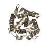
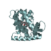
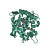
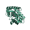
- PDB-7o3u: The crystal structure of obelin from Obelia longissima bound with... -
+ Open data

- Basic information
| Entry | Database: PDB / ID: 7o3u | ||||||
|---|---|---|---|---|---|---|---|
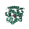
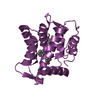
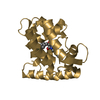
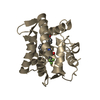
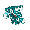
| Title | The crystal structure of obelin from Obelia longissima bound with v-coelenterazine | ||||||
 Components Components | Obelin | ||||||
 Keywords Keywords | LUMINESCENT PROTEIN / bioluminescence / coelenterazine / v-coelenterazine / calcium binding | ||||||
| Function / homology |  Function and homology information Function and homology information | ||||||
| Biological species |  Obelia longissima (invertebrata) Obelia longissima (invertebrata) | ||||||
| Method |  X-RAY DIFFRACTION / SYNCHROTRON / MOLECULAR REPLACEMENT / Resolution: 1.8 Å X-RAY DIFFRACTION / SYNCHROTRON / MOLECULAR REPLACEMENT / Resolution: 1.8 Å | ||||||
 Authors Authors | Larionova, M.D. / Wu, L.J. / Vysotski, E.S. / Liu, Z.-J. | ||||||
 Citation Citation | Journal: Protein Sci. / Year: 2022 Title: Crystal structure of semisynthetic obelin-v. Authors: Larionova, M.D. / Wu, L. / Eremeeva, E.V. / Natashin, P.V. / Gulnov, D.V. / Nemtseva, E.V. / Liu, D. / Liu, Z.J. / Vysotski, E.S. | ||||||
| History |
|
- Structure visualization
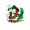
| Structure viewer | Molecule: MolmilJmol/JSmol |
|---|
- Downloads & links
-Download
| PDBx/mmCIF format | 7o3u.cif.gz | 97.6 KB | Display | PDBx/mmCIF format |
|---|---|---|---|---|
| PDB format | pdb7o3u.ent.gz | 74 KB | Display | PDB format |
| PDBx/mmJSON format | 7o3u.json.gz | Tree view | PDBx/mmJSON format | |
| Others |  Other downloads Other downloads |
-Validation report
| Summary document | 7o3u_validation.pdf.gz | 848.5 KB | Display | wwPDB validaton report |
|---|---|---|---|---|
| Full document | 7o3u_full_validation.pdf.gz | 856.5 KB | Display | |
| Data in XML | 7o3u_validation.xml.gz | 11.4 KB | Display | |
| Data in CIF | 7o3u_validation.cif.gz | 15 KB | Display | |
| Arichive directory | https://data.pdbj.org/pub/pdb/validation_reports/o3/7o3uftp://data.pdbj.org/pub/pdb/validation_reports/o3/7o3u | HTTPS FTP |
-Related structure data
| Related structure data |  1qv0S S: Starting model for refinement |
|---|---|
| Similar structure data |
-Links
 PDBj
PDBj- Assembly
| Deposited unit | 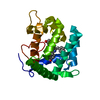
| ||||||||
|---|---|---|---|---|---|---|---|---|---|
| 1 |
| ||||||||
| Unit cell |
| ||||||||
| Components on special symmetry positions |
|
-Components
| #1: Protein | Mass: 22238.904 Da / Num. of mol.: 1 Source method: isolated from a genetically manipulated source Details: v-coelenterazine (v-CTZ or CTZ V) is a synthetic analogue of natural coelenterazine with an additional benzyl ring. Source: (gene. exp.) Obelia longissima (invertebrata) / Plasmid: pET-19b-OL8 / Production host:  |
|---|---|
| #2: Chemical | ChemComp-V1W / 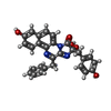  Mass: 479.483 Da / Num. of mol.: 1 / Source method: obtained synthetically / Formula: C28H21N3O5 / Feature type: SUBJECT OF INVESTIGATION Mass: 479.483 Da / Num. of mol.: 1 / Source method: obtained synthetically / Formula: C28H21N3O5 / Feature type: SUBJECT OF INVESTIGATION |
| #3: Chemical | ChemComp-CA /   Mass: 40.078 Da / Num. of mol.: 1 / Source method: obtained synthetically / Formula: Ca Mass: 40.078 Da / Num. of mol.: 1 / Source method: obtained synthetically / Formula: Ca |
| #4: Water | ChemComp-HOH /  Mass: 18.015 Da / Num. of mol.: 86 / Source method: isolated from a natural source / Formula: H2O Mass: 18.015 Da / Num. of mol.: 86 / Source method: isolated from a natural source / Formula: H2O |
| Has ligand of interest | Y |
-Experimental details
-Experiment
| Experiment | Method: X-RAY DIFFRACTION / Number of used crystals: 1 |
|---|
- Sample preparation
| Crystal | Density Matthews: 2.24 Å3/Da / Density % sol: 45.09 % / Description: Light-yellow rod-shaped crystals |
|---|---|
| Crystal grow | Temperature: 277 K / Method: vapor diffusion, hanging drop / pH: 7 / Details: 2.2 M DL-Malic acid |
-Data collection
| Diffraction | Mean temperature: 100 K / Serial crystal experiment: N |
|---|---|
| Diffraction source | Source: SYNCHROTRON / Site: SPring-8  / Beamline: BL45XU / Wavelength: 1 Å / Beamline: BL45XU / Wavelength: 1 Å |
| Detector | Type: DECTRIS PILATUS3 6M / Detector: PIXEL / Date: Feb 9, 2020 |
| Radiation | Protocol: SINGLE WAVELENGTH / Monochromatic (M) / Laue (L): M / Scattering type: x-ray |
| Radiation wavelength | Wavelength: 1 Å / Relative weight: 1 |
| Reflection | Resolution: 1.8→50 Å / Num. obs: 35638 / % possible obs: 99.5 % / Redundancy: 8.3 % / CC1/2: 0.998 / Rmerge(I) obs: 0.099 / Rrim(I) all: 0.106 / Net I/σ(I): 12.77 |
| Reflection shell | Resolution: 1.8→1.91 Å / Redundancy: 5.7 % / Rmerge(I) obs: 0.705 / Mean I/σ(I) obs: 1.86 / Num. unique obs: 5610 / CC1/2: 0.896 / Rrim(I) all: 0.771 / % possible all: 97.2 |
- Processing
| Software |
| |||||||||||||||||||||||||||||||||||||||||||||||||||||||||||||||||||||||||||||||||||||||||||||||||||||||||||||||||||||||||||||||||||||||||||||||||||||||||||||||||||||||||||||||||||||||||||||||||||||||||||||||||||||||||||||||||
|---|---|---|---|---|---|---|---|---|---|---|---|---|---|---|---|---|---|---|---|---|---|---|---|---|---|---|---|---|---|---|---|---|---|---|---|---|---|---|---|---|---|---|---|---|---|---|---|---|---|---|---|---|---|---|---|---|---|---|---|---|---|---|---|---|---|---|---|---|---|---|---|---|---|---|---|---|---|---|---|---|---|---|---|---|---|---|---|---|---|---|---|---|---|---|---|---|---|---|---|---|---|---|---|---|---|---|---|---|---|---|---|---|---|---|---|---|---|---|---|---|---|---|---|---|---|---|---|---|---|---|---|---|---|---|---|---|---|---|---|---|---|---|---|---|---|---|---|---|---|---|---|---|---|---|---|---|---|---|---|---|---|---|---|---|---|---|---|---|---|---|---|---|---|---|---|---|---|---|---|---|---|---|---|---|---|---|---|---|---|---|---|---|---|---|---|---|---|---|---|---|---|---|---|---|---|---|---|---|---|---|---|---|---|---|---|---|---|---|---|---|---|---|---|---|---|---|
| Refinement | Method to determine structure: MOLECULAR REPLACEMENT Starting model: 1qv0 Resolution: 1.8→37.14 Å / SU ML: 0.28 / Cross valid method: THROUGHOUT / σ(F): 1.39 / Phase error: 30.74 / Stereochemistry target values: ML
| |||||||||||||||||||||||||||||||||||||||||||||||||||||||||||||||||||||||||||||||||||||||||||||||||||||||||||||||||||||||||||||||||||||||||||||||||||||||||||||||||||||||||||||||||||||||||||||||||||||||||||||||||||||||||||||||||
| Solvent computation | Shrinkage radii: 0.5 Å / VDW probe radii: 0.9 Å / Solvent model: FLAT BULK SOLVENT MODEL | |||||||||||||||||||||||||||||||||||||||||||||||||||||||||||||||||||||||||||||||||||||||||||||||||||||||||||||||||||||||||||||||||||||||||||||||||||||||||||||||||||||||||||||||||||||||||||||||||||||||||||||||||||||||||||||||||
| Displacement parameters | Biso max: 138.05 Å2 / Biso mean: 56.3241 Å2 / Biso min: 18.77 Å2 | |||||||||||||||||||||||||||||||||||||||||||||||||||||||||||||||||||||||||||||||||||||||||||||||||||||||||||||||||||||||||||||||||||||||||||||||||||||||||||||||||||||||||||||||||||||||||||||||||||||||||||||||||||||||||||||||||
| Refinement step | Cycle: final / Resolution: 1.8→37.14 Å
| |||||||||||||||||||||||||||||||||||||||||||||||||||||||||||||||||||||||||||||||||||||||||||||||||||||||||||||||||||||||||||||||||||||||||||||||||||||||||||||||||||||||||||||||||||||||||||||||||||||||||||||||||||||||||||||||||
| LS refinement shell | Refine-ID: X-RAY DIFFRACTION / Rfactor Rfree error: 0 / Total num. of bins used: 13
| |||||||||||||||||||||||||||||||||||||||||||||||||||||||||||||||||||||||||||||||||||||||||||||||||||||||||||||||||||||||||||||||||||||||||||||||||||||||||||||||||||||||||||||||||||||||||||||||||||||||||||||||||||||||||||||||||
| Refinement TLS params. | Method: refined / Refine-ID: X-RAY DIFFRACTION
| |||||||||||||||||||||||||||||||||||||||||||||||||||||||||||||||||||||||||||||||||||||||||||||||||||||||||||||||||||||||||||||||||||||||||||||||||||||||||||||||||||||||||||||||||||||||||||||||||||||||||||||||||||||||||||||||||
| Refinement TLS group |
|
