 Movie
Movie Controller


+ Open data

- Basic information
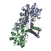
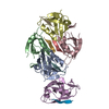
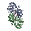
| Entry | Database: PDB / ID: 7o2n | ||||||
|---|---|---|---|---|---|---|---|
| Title | Crystal structure of B. subtilis UGPase YngB | ||||||
 Components Components | Probable UTP--glucose-1-phosphate uridylyltransferase YngB | ||||||
 Keywords Keywords | TRANSFERASE / UTP / glucose-1-phosphate / UDP-glucose | ||||||
| Function / homology |  Function and homology information Function and homology informationenterobacterial common antigen biosynthetic process / UTP-glucose-1-phosphate uridylyltransferase / UTP:glucose-1-phosphate uridylyltransferase activity / UDP-alpha-D-glucose metabolic process / lipopolysaccharide core region biosynthetic process / cytosol Similarity search - Function | ||||||
| Biological species |  | ||||||
| Method |  X-RAY DIFFRACTION / SYNCHROTRON / MOLECULAR REPLACEMENT / Resolution: 2.8 Å X-RAY DIFFRACTION / SYNCHROTRON / MOLECULAR REPLACEMENT / Resolution: 2.8 Å | ||||||
 Authors Authors | Wu, C. | ||||||
| Funding support |  United Kingdom, 1items United Kingdom, 1items
| ||||||
 Citation Citation | Book title: Ph.D.Thesis / Journal: Ph.D.Thesis / Year: 2021 Title: Structural and Functional Characterisation of the Bacillus subtilis Uridylyltransferase YngB Authors: Wu, C. | ||||||
| History |
|
- Structure visualization
| Structure viewer | Molecule: MolmilJmol/JSmol |
|---|
- Downloads & links
-Download
| PDBx/mmCIF format | 7o2n.cif.gz | 123.6 KB | Display | PDBx/mmCIF format |
|---|---|---|---|---|
| PDB format | pdb7o2n.ent.gz | 94.4 KB | Display | PDB format |
| PDBx/mmJSON format | 7o2n.json.gz | Tree view | PDBx/mmJSON format | |
| Others |  Other downloads Other downloads |
-Validation report
| Arichive directory | https://data.pdbj.org/pub/pdb/validation_reports/o2/7o2nftp://data.pdbj.org/pub/pdb/validation_reports/o2/7o2n | HTTPS FTP |
|---|
-Related structure data
| Related structure data |  7b1rS S: Starting model for refinement |
|---|---|
| Similar structure data |


-Links
 PDBj
PDBj

- Assembly
| Deposited unit | 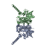
| ||||||||||||||||||
|---|---|---|---|---|---|---|---|---|---|---|---|---|---|---|---|---|---|---|---|
| 1 | 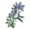
| ||||||||||||||||||
| Unit cell |
| ||||||||||||||||||
| Components on special symmetry positions |
| ||||||||||||||||||
| Noncrystallographic symmetry (NCS) | NCS domain:
NCS domain segments: Component-ID: _ / Ens-ID: 1 / Beg auth comp-ID: LYS / Beg label comp-ID: LYS / End auth comp-ID: LEU / End label comp-ID: LEU / Refine code: _ / Auth seq-ID: 4 - 296 / Label seq-ID: 3 - 295
|
-Components
| #1: Protein | Mass: 33059.949 Da / Num. of mol.: 2 Source method: isolated from a genetically manipulated source Source: (gene. exp.) Strain: 168 / Gene: yngB, BSU18180 / Production host: References: UniProt: O31822, UTP-glucose-1-phosphate uridylyltransferase #2: Water | ChemComp-HOH / |  Mass: 18.015 Da / Num. of mol.: 48 / Source method: isolated from a natural source / Formula: H2O Mass: 18.015 Da / Num. of mol.: 48 / Source method: isolated from a natural source / Formula: H2O |
|---|
-Experimental details
-Experiment
| Experiment | Method: X-RAY DIFFRACTION / Number of used crystals: 1 |
|---|
- Sample preparation
| Crystal | Density Matthews: 2.99 Å3/Da / Density % sol: 58.85 % |
|---|---|
| Crystal grow | Temperature: 289 K / Method: vapor diffusion, sitting drop Details: 0.2M potassium citrate tribasic monohydrate, 0.05M lithium citrate tribasic tetrahydrate, 0.1M sodium phosphate monobasic monohydrate, 21% PEG6000 |
-Data collection
| Diffraction | Mean temperature: 100 K / Serial crystal experiment: N |
|---|---|
| Diffraction source | Source: SYNCHROTRON / Site: Diamond / Beamline: I04 / Wavelength: 0.9795 Å |
| Detector | Type: DECTRIS EIGER2 XE 16M / Detector: PIXEL / Date: Dec 8, 2019 |
| Radiation | Protocol: SINGLE WAVELENGTH / Monochromatic (M) / Laue (L): M / Scattering type: x-ray |
| Radiation wavelength | Wavelength: 0.9795 Å / Relative weight: 1 |
| Reflection | Resolution: 2.8→56.29 Å / Num. obs: 19691 / % possible obs: 100 % / Redundancy: 1.9 % / CC1/2: 0.998 / Rrim(I) all: 0.065 / Net I/σ(I): 9.6 |
| Reflection shell | Resolution: 2.8→2.95 Å / Mean I/σ(I) obs: 2.3 / Num. unique obs: 2814 / CC1/2: 0.91 / Rrim(I) all: 0.397 |
- Processing
| Software |
| |||||||||||||||||||||||||||||||||||||||||||||
|---|---|---|---|---|---|---|---|---|---|---|---|---|---|---|---|---|---|---|---|---|---|---|---|---|---|---|---|---|---|---|---|---|---|---|---|---|---|---|---|---|---|---|---|---|---|---|
| Refinement | Method to determine structure: MOLECULAR REPLACEMENT Starting model: 7B1R Resolution: 2.8→56.29 Å / Cor.coef. Fo:Fc: 0.948 / Cor.coef. Fo:Fc free: 0.924 / SU B: 19.244 / SU ML: 0.349 / Cross valid method: THROUGHOUT / σ(F): 0 / ESU R: 1.176 / ESU R Free: 0.365 / Stereochemistry target values: MAXIMUM LIKELIHOOD / Details: U VALUES : REFINED INDIVIDUALLY
| |||||||||||||||||||||||||||||||||||||||||||||
| Solvent computation | Ion probe radii: 0.8 Å / Shrinkage radii: 0.8 Å / VDW probe radii: 1.2 Å / Solvent model: MASK | |||||||||||||||||||||||||||||||||||||||||||||
| Displacement parameters | Biso max: 179.23 Å2 / Biso mean: 70.184 Å2 / Biso min: 33.55 Å2
| |||||||||||||||||||||||||||||||||||||||||||||
| Refinement step | Cycle: final / Resolution: 2.8→56.29 Å
| |||||||||||||||||||||||||||||||||||||||||||||
| Refine LS restraints |
| |||||||||||||||||||||||||||||||||||||||||||||
| Refine LS restraints NCS | Ens-ID: 1 / Number: 8033 / Refine-ID: X-RAY DIFFRACTION / Type: interatomic distance / Rms dev position: 0.12 Å / Weight position: 0.05
| |||||||||||||||||||||||||||||||||||||||||||||
| LS refinement shell | Resolution: 2.8→2.873 Å / Rfactor Rfree error: 0 / Total num. of bins used: 20
|
