 Movie
Movie Controller


+ Open data

- Basic information
| Entry | Database: PDB / ID: 7nyp | |||||||||
|---|---|---|---|---|---|---|---|---|---|---|
| Title | monomeric acetyl-CoA synthase in closed conformation | |||||||||
 Components Components | CO-methylating acetyl-CoA synthase | |||||||||
 Keywords Keywords | OXIDOREDUCTASE / metalloprotein / nickel / acetogenesis / cluster A / ACS | |||||||||
| Function / homology |  Function and homology information Function and homology informationCO-methylating acetyl-CoA synthase / CO-methylating acetyl-CoA synthase activity / anaerobic carbon-monoxide dehydrogenase activity / acetyl-CoA metabolic process / 4 iron, 4 sulfur cluster binding / metal ion binding Similarity search - Function | |||||||||
| Biological species |   Carboxydothermus hydrogenoformans Z-2901 (bacteria) Carboxydothermus hydrogenoformans Z-2901 (bacteria) | |||||||||
| Method |  X-RAY DIFFRACTION / SYNCHROTRON / MOLECULAR REPLACEMENT / Resolution: 2.1 Å X-RAY DIFFRACTION / SYNCHROTRON / MOLECULAR REPLACEMENT / Resolution: 2.1 Å | |||||||||
 Authors Authors | Kreibich, J. / Jeoung, J.H. / Dobbek, H. | |||||||||
| Funding support |  Germany, 2items Germany, 2items
| |||||||||
 Citation Citation | Journal: To Be Published Title: Ligand binding at the Ni,Ni-[4Fe-4S] cluster of acetyl-CoA synthase Authors: Kreibich, J. / Jeoung, J.H. / Dobbek, H. | |||||||||
| History |
|
- Structure visualization
| Structure viewer | Molecule: MolmilJmol/JSmol |
|---|
- Downloads & links
-Download
| PDBx/mmCIF format | 7nyp.cif.gz | 579.2 KB | Display | PDBx/mmCIF format |
|---|---|---|---|---|
| PDB format | pdb7nyp.ent.gz | 468.3 KB | Display | PDB format |
| PDBx/mmJSON format | 7nyp.json.gz | Tree view | PDBx/mmJSON format | |
| Others |  Other downloads Other downloads |
-Validation report
| Arichive directory | https://data.pdbj.org/pub/pdb/validation_reports/ny/7nypftp://data.pdbj.org/pub/pdb/validation_reports/ny/7nyp | HTTPS FTP |
|---|
-Related structure data
| Related structure data | 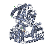 7nysC  7nz5C  7o0dC 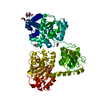 1ru3S S: Starting model for refinement C: citing same article ( |
|---|---|
| Similar structure data |
-Links
 PDBj
PDBj- Assembly
| Deposited unit | 
| ||||||||||||||||||||||||||||||||||||||||||||||||||||||||||||||||||||||||
|---|---|---|---|---|---|---|---|---|---|---|---|---|---|---|---|---|---|---|---|---|---|---|---|---|---|---|---|---|---|---|---|---|---|---|---|---|---|---|---|---|---|---|---|---|---|---|---|---|---|---|---|---|---|---|---|---|---|---|---|---|---|---|---|---|---|---|---|---|---|---|---|---|---|
| 1 | 
| ||||||||||||||||||||||||||||||||||||||||||||||||||||||||||||||||||||||||
| 2 | 
| ||||||||||||||||||||||||||||||||||||||||||||||||||||||||||||||||||||||||
| Unit cell |
| ||||||||||||||||||||||||||||||||||||||||||||||||||||||||||||||||||||||||
| Noncrystallographic symmetry (NCS) | NCS domain:
NCS domain segments:
NCS oper: (Code: givenMatrix: (-0.903122381968, -0.421361284367, -0.0826113262561), (-0.418394305583, 0.90681678123, -0.0512789463184), (0.0965202996504, -0.0117470556616, -0.995261693445)Vector: -34. ...NCS oper: (Code: given Matrix: (-0.903122381968, -0.421361284367, -0.0826113262561), Vector: |
-Components
-Protein , 1 types, 2 molecules AB
| #1: Protein | Mass: 82484.570 Da / Num. of mol.: 2 Source method: isolated from a genetically manipulated source Source: (gene. exp.) Carboxydothermus hydrogenoformans Z-2901 (bacteria)Gene: acsB, CHY_1222 / Plasmid: pQE30 Details (production host): modified vector with twinstrep-tag and TEV cleavage site Production host: References: UniProt: Q3ACS4, CO-methylating acetyl-CoA synthase |
|---|
-Non-polymers , 5 types, 984 molecules 


| #2: Chemical | ChemComp-NI /  Mass: 58.693 Da / Num. of mol.: 4 / Source method: obtained synthetically / Formula: Ni / Feature type: SUBJECT OF INVESTIGATION Mass: 58.693 Da / Num. of mol.: 4 / Source method: obtained synthetically / Formula: Ni / Feature type: SUBJECT OF INVESTIGATION#3: Chemical |  Mass: 351.640 Da / Num. of mol.: 2 / Source method: obtained synthetically / Formula: Fe4S4 Mass: 351.640 Da / Num. of mol.: 2 / Source method: obtained synthetically / Formula: Fe4S4#4: Chemical |  Mass: 106.120 Da / Num. of mol.: 2 / Source method: obtained synthetically / Formula: C4H10O3 Mass: 106.120 Da / Num. of mol.: 2 / Source method: obtained synthetically / Formula: C4H10O3#5: Chemical |  Mass: 618.190 Da / Num. of mol.: 2 / Source method: obtained synthetically / Formula: C18H18O21Ti Mass: 618.190 Da / Num. of mol.: 2 / Source method: obtained synthetically / Formula: C18H18O21Ti#6: Water | ChemComp-HOH / | Mass: 18.015 Da / Num. of mol.: 974 / Source method: isolated from a natural source / Formula: H2O |
|---|
-Details
| Has ligand of interest | Y |
|---|
-Experimental details
-Experiment
| Experiment | Method: X-RAY DIFFRACTION / Number of used crystals: 1 |
|---|
- Sample preparation
| Crystal | Density Matthews: 2.51 Å3/Da / Density % sol: 51.05 % |
|---|---|
| Crystal grow | Temperature: 290 K / Method: vapor diffusion, sitting drop Details: 0.1 M Lithium nitrate, 16% w/v PEG 3350, 5 mM Ti(III)citrate |
-Data collection
| Diffraction | Mean temperature: 100 K / Serial crystal experiment: N |
|---|---|
| Diffraction source | Source: SYNCHROTRON / Site: BESSY / Beamline: 14.1 / Wavelength: 0.918 Å |
| Detector | Type: DECTRIS PILATUS 6M-F / Detector: PIXEL / Date: May 16, 2019 |
| Radiation | Protocol: SINGLE WAVELENGTH / Monochromatic (M) / Laue (L): M / Scattering type: x-ray |
| Radiation wavelength | Wavelength: 0.918 Å / Relative weight: 1 |
| Reflection | Resolution: 2.1→46.44 Å / Num. obs: 96729 / % possible obs: 98.8 % / Redundancy: 2 % / Biso Wilson estimate: 32.07 Å2 / CC1/2: 0.999 / Rmerge(I) obs: 0.039 / Rpim(I) all: 0.039 / Rrim(I) all: 0.056 / Net I/σ(I): 8.6 |
| Reflection shell | Resolution: 2.1→2.18 Å / Rmerge(I) obs: 0.601 / Mean I/σ(I) obs: 1.1 / Num. unique obs: 9478 / CC1/2: 0.585 / Rpim(I) all: 0.601 / Rrim(I) all: 0.849 / % possible all: 97.7 |
- Processing
| Software |
| ||||||||||||||||||||||||||||||||||||||||||||||||||||||||||||||||||||||||||||||||||||||||||||||||||
|---|---|---|---|---|---|---|---|---|---|---|---|---|---|---|---|---|---|---|---|---|---|---|---|---|---|---|---|---|---|---|---|---|---|---|---|---|---|---|---|---|---|---|---|---|---|---|---|---|---|---|---|---|---|---|---|---|---|---|---|---|---|---|---|---|---|---|---|---|---|---|---|---|---|---|---|---|---|---|---|---|---|---|---|---|---|---|---|---|---|---|---|---|---|---|---|---|---|---|---|
| Refinement | Method to determine structure: MOLECULAR REPLACEMENT Starting model: 1RU3 Resolution: 2.1→46.43 Å / SU ML: 0.254 / Cross valid method: FREE R-VALUE / σ(F): 1.33 / Phase error: 27.0482 Stereochemistry target values: GeoStd + Monomer Library + CDL v1.2
| ||||||||||||||||||||||||||||||||||||||||||||||||||||||||||||||||||||||||||||||||||||||||||||||||||
| Solvent computation | Shrinkage radii: 0.9 Å / VDW probe radii: 1.11 Å / Solvent model: FLAT BULK SOLVENT MODEL | ||||||||||||||||||||||||||||||||||||||||||||||||||||||||||||||||||||||||||||||||||||||||||||||||||
| Displacement parameters | Biso mean: 38.11 Å2 | ||||||||||||||||||||||||||||||||||||||||||||||||||||||||||||||||||||||||||||||||||||||||||||||||||
| Refinement step | Cycle: LAST / Resolution: 2.1→46.43 Å
| ||||||||||||||||||||||||||||||||||||||||||||||||||||||||||||||||||||||||||||||||||||||||||||||||||
| Refine LS restraints |
| ||||||||||||||||||||||||||||||||||||||||||||||||||||||||||||||||||||||||||||||||||||||||||||||||||
| Refine LS restraints NCS | Type: Torsion NCS / Rms dev position: 0.887065110097 Å | ||||||||||||||||||||||||||||||||||||||||||||||||||||||||||||||||||||||||||||||||||||||||||||||||||
| LS refinement shell |
|
