Movie
Movie Controller

[English] 日本語
 Yorodumi
- PDB-7ntm: Cryo-EM structure of S.cerevisiae native alcohol dehydrogenase 1 ... -
+ Open data

- Basic information
| Entry | Database: PDB / ID: 7ntm | ||||||
|---|---|---|---|---|---|---|---|
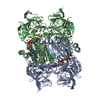
| Title | Cryo-EM structure of S.cerevisiae native alcohol dehydrogenase 1 (ADH1) in its tetrameric apo state | ||||||
 Components Components | Alcohol dehydrogenase 1 | ||||||
 Keywords Keywords | OXIDOREDUCTASE / ADH1 / tetramer / S.cerevisiae | ||||||
| Function / homology |  Function and homology information Function and homology informationmethylglyoxal reductase (NADH) / octanol dehydrogenase (NAD+) activity / methylglyoxal reductase (NADH) activity / pyruvate fermentation to ethanol / : / butanol dehydrogenase (NAD+) activity / ethanol dehydrogenase (NAD+) activity / melatonin binding / alcohol dehydrogenase (NAD+) activity / allyl-alcohol dehydrogenase ...methylglyoxal reductase (NADH) / octanol dehydrogenase (NAD+) activity / methylglyoxal reductase (NADH) activity / pyruvate fermentation to ethanol / : / butanol dehydrogenase (NAD+) activity / ethanol dehydrogenase (NAD+) activity / melatonin binding / alcohol dehydrogenase (NAD+) activity / allyl-alcohol dehydrogenase / allyl-alcohol dehydrogenase activity / alcohol dehydrogenase / zinc ion binding / identical protein binding / plasma membrane / cytoplasm Similarity search - Function | ||||||
| Biological species |  | ||||||
| Method | ELECTRON MICROSCOPY / single particle reconstruction / cryo EM / Resolution: 2.86 Å | ||||||
 Authors Authors | Nzigou Mandouckou, J.A. / Carroni, M. / Haeggstrom, J.Z. / Thulasingam, M. | ||||||
| Funding support |  Sweden, 1items Sweden, 1items
| ||||||
 Citation Citation | Journal: To Be Published Title: Cryo-EM structure of S.cerevisiae native alcohol dehydrogenase 1 (ADH1) in its tetrameric apo state Authors: Nzigou Mandouckou, J.A. / Carroni, M. / Haeggstrom, J.Z. / Thulasingam, M. | ||||||
| History |
|
- Structure visualization
| Structure viewer | Molecule: MolmilJmol/JSmol |
|---|
- Downloads & links
-Download
| PDBx/mmCIF format | 7ntm.cif.gz | 259.8 KB | Display | PDBx/mmCIF format |
|---|---|---|---|---|
| PDB format | pdb7ntm.ent.gz | 207 KB | Display | PDB format |
| PDBx/mmJSON format | 7ntm.json.gz | Tree view | PDBx/mmJSON format | |
| Others |  Other downloads Other downloads |
-Validation report
| Arichive directory | https://data.pdbj.org/pub/pdb/validation_reports/nt/7ntmftp://data.pdbj.org/pub/pdb/validation_reports/nt/7ntm | HTTPS FTP |
|---|
-Related structure data
| Related structure data | 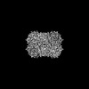 12591MC M: map data used to model this data C: citing same article ( |
|---|---|
| Similar structure data |
-Links
 PDBj
PDBj

- Assembly
| Deposited unit | 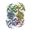
| ||||||||||||||||||||||||||||||||||||||||||||||||||||||||||||||||||||||||||||||||||||||||||||||||||
|---|---|---|---|---|---|---|---|---|---|---|---|---|---|---|---|---|---|---|---|---|---|---|---|---|---|---|---|---|---|---|---|---|---|---|---|---|---|---|---|---|---|---|---|---|---|---|---|---|---|---|---|---|---|---|---|---|---|---|---|---|---|---|---|---|---|---|---|---|---|---|---|---|---|---|---|---|---|---|---|---|---|---|---|---|---|---|---|---|---|---|---|---|---|---|---|---|---|---|---|
| 1 |
| ||||||||||||||||||||||||||||||||||||||||||||||||||||||||||||||||||||||||||||||||||||||||||||||||||
| Noncrystallographic symmetry (NCS) | NCS domain:
NCS domain segments: Component-ID: _ / Beg auth comp-ID: SER / Beg label comp-ID: SER / End auth comp-ID: LYS / End label comp-ID: LYS / Refine code: _ / Auth seq-ID: 1 - 347 / Label seq-ID: 1 - 347
NCS ensembles :
|
-Components
| #1: Protein | Mass: 36759.906 Da / Num. of mol.: 4 / Source method: isolated from a natural source / Source: (natural) #2: Chemical | ChemComp-ZN /   Mass: 65.409 Da / Num. of mol.: 8 / Source method: obtained synthetically / Formula: Zn / Feature type: SUBJECT OF INVESTIGATION Mass: 65.409 Da / Num. of mol.: 8 / Source method: obtained synthetically / Formula: Zn / Feature type: SUBJECT OF INVESTIGATIONHas ligand of interest | Y | Has protein modification | Y | |
|---|
-Experimental details
-Experiment
| Experiment | Method: ELECTRON MICROSCOPY |
|---|---|
| EM experiment | Aggregation state: PARTICLE / 3D reconstruction method: single particle reconstruction |
- Sample preparation
| Component | Name: Tetrameric structure of native ADH1 from S. cereviciae in its apo state. Type: COMPLEX Details: Stable homotetramer, composed of four monomers labelled as A, B, C and D. Entity ID: #1 / Source: NATURAL |
|---|---|
| Molecular weight | Value: 0.147 MDa / Experimental value: YES |
| Source (natural) | Organism: |
| Buffer solution | pH: 7.4 |
| Specimen | Conc.: 2 mg/ml / Embedding applied: NO / Shadowing applied: NO / Staining applied: NO / Vitrification applied: YES |
| Specimen support | Grid material: GOLD / Grid mesh size: 300 divisions/in. / Grid type: Quantifoil R1.2/1.3 |
| Vitrification | Instrument: FEI VITROBOT MARK IV / Cryogen name: ETHANE / Humidity: 100 % / Chamber temperature: 277 K |
- Electron microscopy imaging
| Experimental equipment |  Model: Titan Krios / Image courtesy: FEI Company |
|---|---|
| Microscopy | Model: FEI TITAN KRIOS |
| Electron gun | Electron source:  FIELD EMISSION GUN / Accelerating voltage: 300 kV / Illumination mode: FLOOD BEAM FIELD EMISSION GUN / Accelerating voltage: 300 kV / Illumination mode: FLOOD BEAM |
| Electron lens | Mode: BRIGHT FIELD / Nominal magnification: 130000 X / Nominal defocus max: 3000 nm / Nominal defocus min: 1000 nm / Cs: 2.7 mm / Alignment procedure: COMA FREE |
| Specimen holder | Cryogen: NITROGEN / Specimen holder model: FEI TITAN KRIOS AUTOGRID HOLDER / Residual tilt: 130 mradians |
| Image recording | Average exposure time: 2.5 sec. / Electron dose: 60 e/Å2 / Film or detector model: GATAN K3 BIOQUANTUM (6k x 4k) / Num. of grids imaged: 1 / Num. of real images: 14964 |
| EM imaging optics | Energyfilter name: GIF Bioquantum / Energyfilter slit width: 10 eV |
- Processing
| Software | Name: REFMAC / Version: 5.8.0257 / Classification: refinement | ||||||||||||||||||||||||||||||||||||||||||||||||||||||||||||||||||||||||||||||||||||||||||||||||||||||||||
|---|---|---|---|---|---|---|---|---|---|---|---|---|---|---|---|---|---|---|---|---|---|---|---|---|---|---|---|---|---|---|---|---|---|---|---|---|---|---|---|---|---|---|---|---|---|---|---|---|---|---|---|---|---|---|---|---|---|---|---|---|---|---|---|---|---|---|---|---|---|---|---|---|---|---|---|---|---|---|---|---|---|---|---|---|---|---|---|---|---|---|---|---|---|---|---|---|---|---|---|---|---|---|---|---|---|---|---|
| EM software |
| ||||||||||||||||||||||||||||||||||||||||||||||||||||||||||||||||||||||||||||||||||||||||||||||||||||||||||
| CTF correction | Type: PHASE FLIPPING AND AMPLITUDE CORRECTION | ||||||||||||||||||||||||||||||||||||||||||||||||||||||||||||||||||||||||||||||||||||||||||||||||||||||||||
| Particle selection | Num. of particles selected: 639189 | ||||||||||||||||||||||||||||||||||||||||||||||||||||||||||||||||||||||||||||||||||||||||||||||||||||||||||
| Symmetry | Point symmetry: D2 (2x2 fold dihedral) | ||||||||||||||||||||||||||||||||||||||||||||||||||||||||||||||||||||||||||||||||||||||||||||||||||||||||||
| 3D reconstruction | Resolution: 2.86 Å / Resolution method: FSC 0.143 CUT-OFF / Num. of particles: 222802 / Symmetry type: POINT | ||||||||||||||||||||||||||||||||||||||||||||||||||||||||||||||||||||||||||||||||||||||||||||||||||||||||||
| Atomic model building | B value: 123 / Protocol: BACKBONE TRACE / Space: REAL Details: Model building and refinement were done using using CCP-EM software suite (Burnley et al, 2017). The X-ray structure of yeast ADH1 (PDB: 5ENV) was used as a starting model for model building. ...Details: Model building and refinement were done using using CCP-EM software suite (Burnley et al, 2017). The X-ray structure of yeast ADH1 (PDB: 5ENV) was used as a starting model for model building. This model was docked into our Cryo-EM map using MolRep (Brown et al, 2014). The resulting starting model was manually adjusted in Coot (Emsley et al, 2010). REFMAC 5 (Brown et al, 2014) used for structure model refinement. | ||||||||||||||||||||||||||||||||||||||||||||||||||||||||||||||||||||||||||||||||||||||||||||||||||||||||||
| Atomic model building | PDB-ID: 5ENV Pdb chain-ID: A / Accession code: 5ENV / Pdb chain residue range: 1-347 / Source name: PDB / Type: experimental model | ||||||||||||||||||||||||||||||||||||||||||||||||||||||||||||||||||||||||||||||||||||||||||||||||||||||||||
| Refinement | Resolution: 2.86→242.88 Å / Cor.coef. Fo:Fc: 0.997 / SU B: 0.244 / SU ML: 0.005 / ESU R: 0.012 Stereochemistry target values: MAXIMUM LIKELIHOOD WITH PHASES
| ||||||||||||||||||||||||||||||||||||||||||||||||||||||||||||||||||||||||||||||||||||||||||||||||||||||||||
| Solvent computation | Solvent model: PARAMETERS FOR MASK CACLULATION | ||||||||||||||||||||||||||||||||||||||||||||||||||||||||||||||||||||||||||||||||||||||||||||||||||||||||||
| Displacement parameters | Biso mean: 54.33 Å2
| ||||||||||||||||||||||||||||||||||||||||||||||||||||||||||||||||||||||||||||||||||||||||||||||||||||||||||
| Refinement step | Cycle: 1 / Total: 10336 | ||||||||||||||||||||||||||||||||||||||||||||||||||||||||||||||||||||||||||||||||||||||||||||||||||||||||||
| Refine LS restraints |
|