[English] 日本語
Yorodumi
- PDB-7nsx: Drosophila PGRP-LB wild-type -

+
Open data


ID or keywords:

Loading...

-
Basic information

Entry
Database: PDB / ID: 7nsx
TitleDrosophila PGRP-LB wild-type
ComponentsIsoform A of Peptidoglycan-recognition protein LB
Keywords
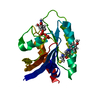
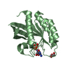
IMMUNE SYSTEM / Peptidoglycan recognition protein / PGRP / PGRP-LB / Drosophila
Function / homology
Function and homology information


negative regulation of biosynthetic process of antibacterial peptides active against Gram-negative bacteria / negative regulation of peptidoglycan recognition protein signaling pathway / N-acetylmuramoyl-L-alanine amidase / N-acetylmuramoyl-L-alanine amidase activity / peptidoglycan binding / peptidoglycan catabolic process / defense response to Gram-positive bacterium / innate immune response / zinc ion binding / extracellular region
Similarity search - Function
Peptidoglycan recognition protein, PGRP-S / Peptidoglycan recognition protein family domain, metazoa/bacteria / Peptidoglycan recognition protein / Animal peptidoglycan recognition proteins homologous to Bacteriophage T3 lysozyme. / Ami_2 / N-acetylmuramoyl-L-alanine amidase / N-acetylmuramoyl-L-alanine amidase domain / N-acetylmuramoyl-L-alanine amidase/PGRP domain superfamily
Similarity search - Domain/homology
Peptidoglycan-recognition protein LB
Similarity search - Component
Biological speciesDrosophila melanogaster (fruit fly)
MethodX-RAY DIFFRACTION / SYNCHROTRON / MOLECULAR REPLACEMENT / Resolution: 1.9 Å
AuthorsOrlans, J. / Aller, P. / Da Silva, P.
CitationJournal: Int J Mol Sci / Year: 2021
Title: PGRP-LB: An Inside View into the Mechanism of the Amidase Reaction.
Authors: Orlans, J. / Vincent-Monegat, C. / Rahioui, I. / Sivignon, C. / Butryn, A. / Soulere, L. / Zaidman-Remy, A. / Orville, A.M. / Heddi, A. / Aller, P. / Da Silva, P.
History
DepositionMar 8, 2021Deposition site: PDBE / Processing site: PDBE
Revision 1.0May 19, 2021Provider: repository / Type: Initial release
Revision 1.1Jun 16, 2021Group: Database references / Category: citation
Item: _citation.journal_volume / _citation.pdbx_database_id_PubMed / _citation.title
Revision 1.2Jan 31, 2024Group: Data collection / Database references ...Data collection / Database references / Derived calculations / Refinement description
Category: atom_type / chem_comp_atom ...atom_type / chem_comp_atom / chem_comp_bond / database_2 / pdbx_initial_refinement_model
Item: _atom_type.pdbx_N_electrons / _atom_type.pdbx_scat_Z ..._atom_type.pdbx_N_electrons / _atom_type.pdbx_scat_Z / _database_2.pdbx_DOI / _database_2.pdbx_database_accession

-
Structure visualization

Structure viewerMolecule:
MolmilJmol/JSmol

Downloads & links

-
Assembly

Deposited unit
AAA: Isoform A of Peptidoglycan-recognition protein LB
hetero molecules


Theoretical massNumber of molelcules
Total (without water)23,8522
Polymers23,7871
Non-polymers651
Water91951
1


  • Idetical with deposited unit
  • defined by author&software
TypeNameSymmetry operationNumber
identity operation1_555x,y,z1
Buried area100 Å2
ΔGint-37 kcal/mol
Surface area8090 Å2
MethodPISA
Unit cell
Length a, b, c (Å)39.869, 70.546, 112.828
Angle α, β, γ (deg.)90.000, 90.000, 90.000
Int Tables number20
Space group name H-MC2221

-
Components

#1: Protein Isoform A of Peptidoglycan-recognition protein LB / PGRP-LB


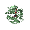
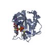
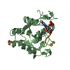
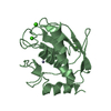
Mass: 23787.029 Da / Num. of mol.: 1
Source method: isolated from a genetically manipulated source
Details: Please can you count the first two residue as -1 and -2 because they belong to the cleavage site of the tag. The number 1 of the sequence would be third residue.
Source: (gene. exp.) Drosophila melanogaster (fruit fly) / Gene: PGRP-LB, CG14704 / Production host: Escherichia coli BL21(DE3) (bacteria)
References: UniProt: Q8INK6, N-acetylmuramoyl-L-alanine amidase
#2: Chemical ChemComp-ZN / ZINC ION


Mass: 65.409 Da / Num. of mol.: 1 / Source method: obtained synthetically / Formula: Zn / Feature type: SUBJECT OF INVESTIGATION
#3: Water ChemComp-HOH / water


Mass: 18.015 Da / Num. of mol.: 51 / Source method: isolated from a natural source / Formula: H2O
Has ligand of interestY

-
Experimental details

-
Experiment

ExperimentMethod: X-RAY DIFFRACTION / Number of used crystals: 1

-
Sample preparation

CrystalDensity Matthews: 1.67 Å3/Da / Density % sol: 26.24 %
Crystal growTemperature: 293 K / Method: vapor diffusion, sitting drop / pH: 7 / Details: 0.1M HEPES pH7.0, 30% v/v Jeffamine ED-2003

-
Data collection

DiffractionMean temperature: 100 K / Serial crystal experiment: N
Diffraction sourceSource: SYNCHROTRON / Site: Diamond / Beamline: I24 / Wavelength: 0.969 Å
DetectorType: DECTRIS PILATUS3 6M / Detector: PIXEL / Date: Sep 27, 2018
RadiationMonochromator: Double crystals monochromator, Si111 / Protocol: SINGLE WAVELENGTH / Monochromatic (M) / Laue (L): M / Scattering type: x-ray
Radiation wavelengthWavelength: 0.969 Å / Relative weight: 1
ReflectionResolution: 1.9→112.83 Å / Num. obs: 12942 / % possible obs: 99.8 % / Redundancy: 7.9 % / CC1/2: 0.993 / Rmerge(I) obs: 0.133 / Rpim(I) all: 0.073 / Rrim(I) all: 0.153 / Net I/σ(I): 8.5
Reflection shell

Diffraction-ID: 1

Resolution (Å)Redundancy (%)Rmerge(I) obsNum. unique obsCC1/2Rpim(I) allRrim(I) all
9.11-112.8360.0431500.9980.0240.049
1.9-1.947.21.1978080.5620.6991.394

-
Processing

Software
NameVersionClassification
REFMAC5.8.0267refinement
DIALSdata processing
Aimless0.7.4data reduction
Cootmodel building
GDAdata collection
Aimlessdata scaling
PHASERphasing
RefinementMethod to determine structure: MOLECULAR REPLACEMENT
Starting model: 1OHT
Resolution: 1.9→56.478 Å / Cor.coef. Fo:Fc: 0.963 / Cor.coef. Fo:Fc free: 0.944 / SU B: 3.89 / SU ML: 0.109 / Cross valid method: FREE R-VALUE / ESU R: 0.159 / ESU R Free: 0.139
RfactorNum. reflection% reflection
Rfree0.2128 680 5.278 %
Rwork0.1799 12203 -
all0.182 --
obs-12883 99.667 %
Solvent computationIon probe radii: 0.8 Å / Shrinkage radii: 0.8 Å / VDW probe radii: 1.2 Å / Solvent model: MASK BULK SOLVENT
Displacement parametersBiso mean: 27.154 Å2
Baniso -1Baniso -2Baniso -3
1--0.309 Å20 Å2-0 Å2
2--0.223 Å20 Å2
3---0.086 Å2
Refinement stepCycle: LAST / Resolution: 1.9→56.478 Å
ProteinNucleic acidLigandSolventTotal
Num. atoms1334 0 1 51 1386
Refine LS restraints
Refine-IDTypeDev idealDev ideal targetNumber
X-RAY DIFFRACTIONr_bond_refined_d0.010.0131376
X-RAY DIFFRACTIONr_bond_other_d0.0010.0171241
X-RAY DIFFRACTIONr_angle_refined_deg1.5971.6441869
X-RAY DIFFRACTIONr_angle_other_deg1.3681.5812856
X-RAY DIFFRACTIONr_dihedral_angle_1_deg6.9125169
X-RAY DIFFRACTIONr_dihedral_angle_2_deg34.27420.94674
X-RAY DIFFRACTIONr_dihedral_angle_3_deg13.19915210
X-RAY DIFFRACTIONr_dihedral_angle_4_deg16.6531510
X-RAY DIFFRACTIONr_chiral_restr0.0740.2171
X-RAY DIFFRACTIONr_gen_planes_refined0.0080.021581
X-RAY DIFFRACTIONr_gen_planes_other0.0010.02335
X-RAY DIFFRACTIONr_nbd_refined0.2030.2269
X-RAY DIFFRACTIONr_symmetry_nbd_other0.1850.21190
X-RAY DIFFRACTIONr_nbtor_refined0.1730.2669
X-RAY DIFFRACTIONr_symmetry_nbtor_other0.0770.2626
X-RAY DIFFRACTIONr_xyhbond_nbd_refined0.1410.247
X-RAY DIFFRACTIONr_metal_ion_refined0.0220.21
X-RAY DIFFRACTIONr_symmetry_nbd_refined0.2390.213
X-RAY DIFFRACTIONr_nbd_other0.2030.238
X-RAY DIFFRACTIONr_symmetry_xyhbond_nbd_refined0.1190.26
X-RAY DIFFRACTIONr_mcbond_it2.2242.683679
X-RAY DIFFRACTIONr_mcbond_other2.2072.677678
X-RAY DIFFRACTIONr_mcangle_it3.1164.009847
X-RAY DIFFRACTIONr_mcangle_other3.1254.014848
X-RAY DIFFRACTIONr_scbond_it2.9493.061697
X-RAY DIFFRACTIONr_scbond_other2.9143.059697
X-RAY DIFFRACTIONr_scangle_it4.4314.4741022
X-RAY DIFFRACTIONr_scangle_other4.4034.4731022
X-RAY DIFFRACTIONr_lrange_it5.56631.4241531
X-RAY DIFFRACTIONr_lrange_other5.56231.3471526
LS refinement shell
Resolution (Å)Rfactor RfreeNum. reflection RfreeRfactor RworkNum. reflection RworkRefine-ID% reflection obs (%)
1.9-1.9490.333360.279881X-RAY DIFFRACTION99.1351
1.949-2.0030.307670.248853X-RAY DIFFRACTION99.5671
2.003-2.0610.305410.218833X-RAY DIFFRACTION99.4312
2.061-2.1240.247330.217843X-RAY DIFFRACTION99.6587
2.124-2.1940.259520.209763X-RAY DIFFRACTION99.2692
2.194-2.2710.227600.199761X-RAY DIFFRACTION99.2745
2.271-2.3570.193390.19752X-RAY DIFFRACTION99.6222
2.357-2.4530.337330.188711X-RAY DIFFRACTION99.8658
2.453-2.5620.243300.189710X-RAY DIFFRACTION99.8651
2.562-2.6870.226390.187657X-RAY DIFFRACTION99.5708
2.687-2.8320.244440.173621X-RAY DIFFRACTION100
2.832-3.0040.223370.18597X-RAY DIFFRACTION100
3.004-3.2110.17280.17561X-RAY DIFFRACTION100
3.211-3.4680.14250.16536X-RAY DIFFRACTION100
3.468-3.7990.187330.161481X-RAY DIFFRACTION100
3.799-4.2460.207240.151448X-RAY DIFFRACTION100
4.246-4.9020.171230.143404X-RAY DIFFRACTION100
4.902-6.0010.1990.173357X-RAY DIFFRACTION100
6.001-8.4750.199130.198271X-RAY DIFFRACTION99.6491
8.475-56.4780.143140.15163X-RAY DIFFRACTION98.8827

+
About Yorodumi

-
News

-
Feb 9, 2022. New format data for meta-information of EMDB entries

New format data for meta-information of EMDB entries

  • Version 3 of the EMDB header file is now the official format.
  • The previous official version 1.9 will be removed from the archive.

Related info.:EMDB header

External links:wwPDB to switch to version 3 of the EMDB data model

-
Aug 12, 2020. Covid-19 info

Covid-19 info

URL: https://pdbjlvh1.pdbj.org/emnavi/covid19.php

New page: Covid-19 featured information page in EM Navigator.

Related info.:Covid-19 info / Mar 5, 2020. Novel coronavirus structure data

+
Mar 5, 2020. Novel coronavirus structure data

Novel coronavirus structure data

Related info.:Yorodumi Speices / Aug 12, 2020. Covid-19 info

External links:COVID-19 featured content - PDBj / Molecule of the Month (242):Coronavirus Proteases

+
Jan 31, 2019. EMDB accession codes are about to change! (news from PDBe EMDB page)

EMDB accession codes are about to change! (news from PDBe EMDB page)

  • The allocation of 4 digits for EMDB accession codes will soon come to an end. Whilst these codes will remain in use, new EMDB accession codes will include an additional digit and will expand incrementally as the available range of codes is exhausted. The current 4-digit format prefixed with “EMD-” (i.e. EMD-XXXX) will advance to a 5-digit format (i.e. EMD-XXXXX), and so on. It is currently estimated that the 4-digit codes will be depleted around Spring 2019, at which point the 5-digit format will come into force.
  • The EM Navigator/Yorodumi systems omit the EMD- prefix.

Related info.:Q: What is EMD? / ID/Accession-code notation in Yorodumi/EM Navigator

External links:EMDB Accession Codes are Changing Soon! / Contact to PDBj

+
Jul 12, 2017. Major update of PDB

Major update of PDB

  • wwPDB released updated PDB data conforming to the new PDBx/mmCIF dictionary.
  • This is a major update changing the version number from 4 to 5, and with Remediation, in which all the entries are updated.
  • In this update, many items about electron microscopy experimental information are reorganized (e.g. em_software).
  • Now, EM Navigator and Yorodumi are based on the updated data.

External links:wwPDB Remediation / Enriched Model Files Conforming to OneDep Data Standards Now Available in the PDB FTP Archive

-
Yorodumi

Thousand views of thousand structures

  • Yorodumi is a browser for structure data from EMDB, PDB, SASBDB, etc.
  • This page is also the successor to EM Navigator detail page, and also detail information page/front-end page for Omokage search.
  • The word "yorodu" (or yorozu) is an old Japanese word meaning "ten thousand". "mi" (miru) is to see.

Related info.:EMDB / PDB / SASBDB / Comparison of 3 databanks / Yorodumi Search / Aug 31, 2016. New EM Navigator & Yorodumi / Yorodumi Papers / Jmol/JSmol / Function and homology information / Changes in new EM Navigator and Yorodumi

Read more