 Movie
Movie Controller

[English] 日本語
 Yorodumi
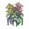
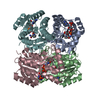
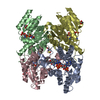
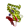
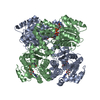
- PDB-7nm8: The crystal structure of the antimycin pathway standalone ketored... -
+ Open data

- Basic information
| Entry | Database: PDB / ID: 7nm8 | ||||||
|---|---|---|---|---|---|---|---|
| Title | The crystal structure of the antimycin pathway standalone ketoreductase, AntM, in complex with NADPH | ||||||
 Components Components | Antimycin pathway standalone ketoreductase, AntM | ||||||
 Keywords Keywords | OXIDOREDUCTASE / Ketoreductase / Standalone / Antimycin / NADPH | ||||||
| Function / homology | Chem-NDP Function and homology information Function and homology information | ||||||
| Biological species |  Streptomyces albus (bacteria) Streptomyces albus (bacteria) | ||||||
| Method |  X-RAY DIFFRACTION / SYNCHROTRON / MOLECULAR REPLACEMENT / Resolution: 1.7 Å X-RAY DIFFRACTION / SYNCHROTRON / MOLECULAR REPLACEMENT / Resolution: 1.7 Å | ||||||
 Authors Authors | Fazal, A. / Hemsworth, G.R. / Webb, M.E. / Seipke, R.F. | ||||||
| Funding support |  United Kingdom, 1items United Kingdom, 1items
| ||||||
 Citation Citation | Journal: Acs Chem.Biol. / Year: 2021 Title: A Standalone beta-Ketoreductase Acts Concomitantly with Biosynthesis of the Antimycin Scaffold. Authors: Fazal, A. / Hemsworth, G.R. / Webb, M.E. / Seipke, R.F. | ||||||
| History |
|
- Structure visualization
| Structure viewer | Molecule: MolmilJmol/JSmol |
|---|
- Downloads & links
-Download
| PDBx/mmCIF format | 7nm8.cif.gz | 101.1 KB | Display | PDBx/mmCIF format |
|---|---|---|---|---|
| PDB format | pdb7nm8.ent.gz | Display | PDB format | |
| PDBx/mmJSON format | 7nm8.json.gz | Tree view | PDBx/mmJSON format | |
| Others |  Other downloads Other downloads |
-Validation report
| Summary document | 7nm8_validation.pdf.gz | 820.3 KB | Display | wwPDB validaton report |
|---|---|---|---|---|
| Full document | 7nm8_full_validation.pdf.gz | 821.9 KB | Display | |
| Data in XML | 7nm8_validation.xml.gz | 12.1 KB | Display | |
| Data in CIF | 7nm8_validation.cif.gz | 16.4 KB | Display | |
| Arichive directory | https://data.pdbj.org/pub/pdb/validation_reports/nm/7nm8ftp://data.pdbj.org/pub/pdb/validation_reports/nm/7nm8 | HTTPS FTP |
-Related structure data
| Related structure data |  7nm7C  4ospS S: Starting model for refinement C: citing same article ( |
|---|---|
| Similar structure data |


-Links
 PDBj
PDBj

- Assembly
| Deposited unit | 
| ||||||||
|---|---|---|---|---|---|---|---|---|---|
| 1 | 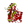
| ||||||||
| Unit cell |
| ||||||||
| Components on special symmetry positions |
|
-Components
| #1: Protein | Mass: 30024.676 Da / Num. of mol.: 1 Source method: isolated from a genetically manipulated source Source: (gene. exp.) Streptomyces albus (bacteria) / Strain: S4 / Gene: antM / Production host: |
|---|---|
| #2: Chemical | ChemComp-NDP /   Mass: 745.421 Da / Num. of mol.: 1 / Source method: obtained synthetically / Formula: C21H30N7O17P3 / Feature type: SUBJECT OF INVESTIGATION Mass: 745.421 Da / Num. of mol.: 1 / Source method: obtained synthetically / Formula: C21H30N7O17P3 / Feature type: SUBJECT OF INVESTIGATION |
| #3: Chemical | ChemComp-GOL /   Mass: 92.094 Da / Num. of mol.: 1 / Source method: obtained synthetically / Formula: C3H8O3 Mass: 92.094 Da / Num. of mol.: 1 / Source method: obtained synthetically / Formula: C3H8O3 |
| #4: Water | ChemComp-HOH /  Mass: 18.015 Da / Num. of mol.: 75 / Source method: isolated from a natural source / Formula: H2O Mass: 18.015 Da / Num. of mol.: 75 / Source method: isolated from a natural source / Formula: H2O |
| Has ligand of interest | Y |
-Experimental details
-Experiment
| Experiment | Method: X-RAY DIFFRACTION / Number of used crystals: 1 |
|---|
- Sample preparation
| Crystal | Density Matthews: 2.33 Å3/Da / Density % sol: 47.11 % |
|---|---|
| Crystal grow | Temperature: 298.15 K / Method: vapor diffusion, sitting drop Details: 0.1 M Tris, pH 8.5, 5% w/v PEG 8K, 20% v/v PEG 300, 10% v/v glycerol |
-Data collection
| Diffraction | Mean temperature: 100 K / Serial crystal experiment: N |
|---|---|
| Diffraction source | Source: SYNCHROTRON / Site: Diamond / Beamline: I04 / Wavelength: 0.9795 Å |
| Detector | Type: DECTRIS EIGER2 X 16M / Detector: PIXEL / Date: Oct 14, 2018 |
| Radiation | Protocol: SINGLE WAVELENGTH / Monochromatic (M) / Laue (L): M / Scattering type: x-ray |
| Radiation wavelength | Wavelength: 0.9795 Å / Relative weight: 1 |
| Reflection | Resolution: 1.7→65.36 Å / Num. obs: 33683 / % possible obs: 100 % / Redundancy: 21.2 % / CC1/2: 1 / Net I/σ(I): 24.7 |
| Reflection shell | Resolution: 1.7→1.73 Å / Num. unique obs: 1652 / CC1/2: 0.958 |
- Processing
| Software |
| |||||||||||||||||||||||||||||||||||||||||||||||||||||||||||||||||||||||||||||||||||||||||||||||||||||||||||||||||||||||||||||||||||||||||||||||||||||||||||
|---|---|---|---|---|---|---|---|---|---|---|---|---|---|---|---|---|---|---|---|---|---|---|---|---|---|---|---|---|---|---|---|---|---|---|---|---|---|---|---|---|---|---|---|---|---|---|---|---|---|---|---|---|---|---|---|---|---|---|---|---|---|---|---|---|---|---|---|---|---|---|---|---|---|---|---|---|---|---|---|---|---|---|---|---|---|---|---|---|---|---|---|---|---|---|---|---|---|---|---|---|---|---|---|---|---|---|---|---|---|---|---|---|---|---|---|---|---|---|---|---|---|---|---|---|---|---|---|---|---|---|---|---|---|---|---|---|---|---|---|---|---|---|---|---|---|---|---|---|---|---|---|---|---|---|---|---|
| Refinement | Method to determine structure: MOLECULAR REPLACEMENT Starting model: 4osp Resolution: 1.7→65.36 Å / Cor.coef. Fo:Fc: 0.966 / Cor.coef. Fo:Fc free: 0.96 / Cross valid method: THROUGHOUT / ESU R: 0.101 / ESU R Free: 0.098 Details: Hydrogens have been added in their riding positions
| |||||||||||||||||||||||||||||||||||||||||||||||||||||||||||||||||||||||||||||||||||||||||||||||||||||||||||||||||||||||||||||||||||||||||||||||||||||||||||
| Solvent computation | Ion probe radii: 0.8 Å / Shrinkage radii: 0.8 Å / VDW probe radii: 1.2 Å / Solvent model: MASK BULK SOLVENT | |||||||||||||||||||||||||||||||||||||||||||||||||||||||||||||||||||||||||||||||||||||||||||||||||||||||||||||||||||||||||||||||||||||||||||||||||||||||||||
| Displacement parameters | Biso mean: 38.772 Å2
| |||||||||||||||||||||||||||||||||||||||||||||||||||||||||||||||||||||||||||||||||||||||||||||||||||||||||||||||||||||||||||||||||||||||||||||||||||||||||||
| Refinement step | Cycle: LAST / Resolution: 1.7→65.36 Å
| |||||||||||||||||||||||||||||||||||||||||||||||||||||||||||||||||||||||||||||||||||||||||||||||||||||||||||||||||||||||||||||||||||||||||||||||||||||||||||
| Refine LS restraints |
| |||||||||||||||||||||||||||||||||||||||||||||||||||||||||||||||||||||||||||||||||||||||||||||||||||||||||||||||||||||||||||||||||||||||||||||||||||||||||||
| LS refinement shell |
|
