 Movie
Movie Controller

[English] 日本語
 Yorodumi
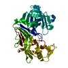
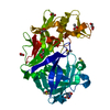
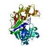
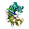
- PDB-7nkw: Endothiapepsin structure obtained at 298K after a soaking with fr... -
+ Open data

- Basic information
| Entry | Database: PDB / ID: 7nkw | ||||||
|---|---|---|---|---|---|---|---|
| Title | Endothiapepsin structure obtained at 298K after a soaking with fragment JFD03909 from a dataset collected with JUNGFRAU detector | ||||||
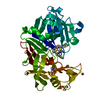
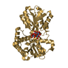
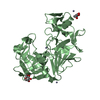
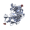
 Components Components | Endothiapepsin | ||||||
 Keywords Keywords | HYDROLASE / FBDD / room temperature / JUNGFRAU | ||||||
| Function / homology |  Function and homology information Function and homology information | ||||||
| Biological species |  Cryphonectria parasitica (chestnut blight fungus) Cryphonectria parasitica (chestnut blight fungus) | ||||||
| Method |  X-RAY DIFFRACTION / SYNCHROTRON / MOLECULAR REPLACEMENT / molecular replacement / Resolution: 2.27 Å X-RAY DIFFRACTION / SYNCHROTRON / MOLECULAR REPLACEMENT / molecular replacement / Resolution: 2.27 Å | ||||||
 Authors Authors | Engilberge, S. / Huang, C.-Y. / Leonarski, F. / Wojdyla, J.A. / Marsh, M. / Olieric, V. / Wang, M. | ||||||
| Funding support |  Switzerland, 1items Switzerland, 1items
| ||||||
 Citation Citation | Journal: To Be Published Title: Endothiapepsin structure obtained at 298K after a soaking with fragment JFD03909 from a dataset collected with JUNGFRAU detector Authors: Engilberge, S. / Huang, C.-Y. / Smith, K.M.L. / Eris, D. / Wojdyla, J.A. / Olieric, V. / Leonarski, F. / Sharpe, M. / Wang, M. | ||||||
| History |
|
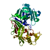
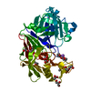
- Structure visualization
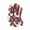
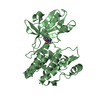
| Structure viewer | Molecule: MolmilJmol/JSmol |
|---|
- Downloads & links
-Download
| PDBx/mmCIF format | 7nkw.cif.gz | 133.4 KB | Display | PDBx/mmCIF format |
|---|---|---|---|---|
| PDB format | pdb7nkw.ent.gz | 103 KB | Display | PDB format |
| PDBx/mmJSON format | 7nkw.json.gz | Tree view | PDBx/mmJSON format | |
| Others |  Other downloads Other downloads |
-Validation report
| Summary document | 7nkw_validation.pdf.gz | 428.8 KB | Display | wwPDB validaton report |
|---|---|---|---|---|
| Full document | 7nkw_full_validation.pdf.gz | 429.5 KB | Display | |
| Data in XML | 7nkw_validation.xml.gz | 13.3 KB | Display | |
| Data in CIF | 7nkw_validation.cif.gz | 18.4 KB | Display | |
| Arichive directory | https://data.pdbj.org/pub/pdb/validation_reports/nk/7nkwftp://data.pdbj.org/pub/pdb/validation_reports/nk/7nkw | HTTPS FTP |
-Related structure data
| Related structure data |  6rsvS S: Starting model for refinement |
|---|---|
| Similar structure data |


-Links
 PDBj
PDBj
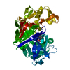
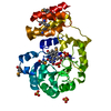
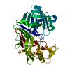
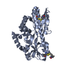
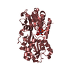
- Assembly
| Deposited unit | 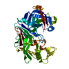
| ||||||||
|---|---|---|---|---|---|---|---|---|---|
| 1 |
| ||||||||
| Unit cell |
|
-Components
| #1: Protein | Mass: 33813.855 Da / Num. of mol.: 1 / Source method: isolated from a natural source Source: (natural) Cryphonectria parasitica (chestnut blight fungus)References: UniProt: P11838, endothiapepsin |
|---|---|
| #2: Chemical | ChemComp-DMS /   Mass: 78.133 Da / Num. of mol.: 1 / Source method: obtained synthetically / Formula: C2H6OS / Comment: DMSO, precipitant*YM Mass: 78.133 Da / Num. of mol.: 1 / Source method: obtained synthetically / Formula: C2H6OS / Comment: DMSO, precipitant*YM |
| #3: Water | ChemComp-HOH /  Mass: 18.015 Da / Num. of mol.: 85 / Source method: isolated from a natural source / Formula: H2O Mass: 18.015 Da / Num. of mol.: 85 / Source method: isolated from a natural source / Formula: H2O |
| Has protein modification | Y |
-Experimental details
-Experiment
| Experiment | Method: X-RAY DIFFRACTION / Number of used crystals: 1 |
|---|
- Sample preparation
| Crystal | Density Matthews: 2.55 Å3/Da / Density % sol: 51.67 % |
|---|---|
| Crystal grow | Temperature: 293 K / Method: vapor diffusion Details: 100 mM ammonium acetate, 100 mM sodium acetate pH 4.6 and 26 to 30% PEG 4000 |
-Data collection
| Diffraction | Mean temperature: 100 K / Serial crystal experiment: N |
|---|---|
| Diffraction source | Source: SYNCHROTRON / Site: SLS / Beamline: X06DA / Wavelength: 1 Å |
| Detector | Type: PSI JUNGFRAU 4M / Detector: PIXEL / Date: Oct 9, 2020 |
| Radiation | Protocol: SINGLE WAVELENGTH / Monochromatic (M) / Laue (L): M / Scattering type: x-ray |
| Radiation wavelength | Wavelength: 1 Å / Relative weight: 1 |
| Reflection | Resolution: 2.27→43.66 Å / Num. obs: 15626 / % possible obs: 99.31 % / Redundancy: 5.8 % / Biso Wilson estimate: 44.94 Å2 / CC1/2: 0.96 / Rpim(I) all: 0.23 / Net I/σ(I): 3.45 |
| Reflection shell | Resolution: 2.27→2.35 Å / Mean I/σ(I) obs: 0.58 / Num. unique obs: 1487 / CC1/2: 0.09 / Rpim(I) all: 1.79 |
-Phasing
| Phasing | Method: molecular replacement |
|---|
- Processing
| Software |
| ||||||||||||||||||||||||||||||||||||||||||||||||||||||||||||||||||||||||||||||||||||||||||||||||||||||||||||
|---|---|---|---|---|---|---|---|---|---|---|---|---|---|---|---|---|---|---|---|---|---|---|---|---|---|---|---|---|---|---|---|---|---|---|---|---|---|---|---|---|---|---|---|---|---|---|---|---|---|---|---|---|---|---|---|---|---|---|---|---|---|---|---|---|---|---|---|---|---|---|---|---|---|---|---|---|---|---|---|---|---|---|---|---|---|---|---|---|---|---|---|---|---|---|---|---|---|---|---|---|---|---|---|---|---|---|---|---|---|
| Refinement | Method to determine structure: MOLECULAR REPLACEMENT Starting model: 6RSV Resolution: 2.27→43.66 Å / Cor.coef. Fo:Fc: 0.944 / Cor.coef. Fo:Fc free: 0.934 / Cross valid method: THROUGHOUT / σ(F): 0 / SU R Blow DPI: 0.266 / SU Rfree Blow DPI: 0.2
| ||||||||||||||||||||||||||||||||||||||||||||||||||||||||||||||||||||||||||||||||||||||||||||||||||||||||||||
| Displacement parameters | Biso max: 122.53 Å2 / Biso mean: 54.94 Å2 / Biso min: 22.32 Å2
| ||||||||||||||||||||||||||||||||||||||||||||||||||||||||||||||||||||||||||||||||||||||||||||||||||||||||||||
| Refine analyze | Luzzati coordinate error obs: 0.35 Å | ||||||||||||||||||||||||||||||||||||||||||||||||||||||||||||||||||||||||||||||||||||||||||||||||||||||||||||
| Refinement step | Cycle: final / Resolution: 2.27→43.66 Å
| ||||||||||||||||||||||||||||||||||||||||||||||||||||||||||||||||||||||||||||||||||||||||||||||||||||||||||||
| Refine LS restraints |
| ||||||||||||||||||||||||||||||||||||||||||||||||||||||||||||||||||||||||||||||||||||||||||||||||||||||||||||
| LS refinement shell | Resolution: 2.27→2.29 Å / Rfactor Rfree error: 0 / Total num. of bins used: 39
| ||||||||||||||||||||||||||||||||||||||||||||||||||||||||||||||||||||||||||||||||||||||||||||||||||||||||||||
| Refinement TLS params. | Method: refined / Origin x: 4.3637 Å / Origin y: 0.6876 Å / Origin z: 5.6119 Å
| ||||||||||||||||||||||||||||||||||||||||||||||||||||||||||||||||||||||||||||||||||||||||||||||||||||||||||||
| Refinement TLS group | Selection details: { A|* } |
