[English] 日本語
Yorodumi
- PDB-7ng0: Crystal structure of N- and C-terminally truncated Geobacillus th... -

+
Open data


ID or keywords:

Loading...

-
Basic information

Entry
Database: PDB / ID: 7ng0
TitleCrystal structure of N- and C-terminally truncated Geobacillus thermoleovorans nucleoid occlusion protein Noc
ComponentsNucleoid occlusion protein
KeywordsDNA BINDING PROTEIN / CTP switch / chromosome segregation / protein-DNA recognition / peripheral membrane protein
Function / homology
Function and homology information


nucleoid / division septum assembly / DNA binding / cytoplasm
Similarity search - Function
Nucleoid occlusion protein / ParB/Spo0J, HTH domain / HTH domain found in ParB protein / ParB/RepB/Spo0J partition protein / ParB/Sulfiredoxin domain / ParB/Sulfiredoxin / ParB-like nuclease domain / ParB/Sulfiredoxin superfamily
Similarity search - Domain/homology
Nucleoid occlusion protein
Similarity search - Component
Biological speciesGeobacillus thermoleovorans CCB_US3_UF5 (bacteria)
MethodX-RAY DIFFRACTION / SYNCHROTRON / MOLECULAR REPLACEMENT / Resolution: 2.95 Å
AuthorsJalal, A.S.B. / Tran, N.T. / Wu, L.J. / Ramakrishnan, K. / Rejzek, M. / Stevenson, C.E.M. / Lawson, D.M. / Errington, J. / Le, T.B.K.
Funding support United Kingdom, 4items
OrganizationGrant numberCountry
Royal SocietyURF-R-201020 United Kingdom
Wellcome Trust209500 United Kingdom
Royal SocietyRG150448 United Kingdom
Biotechnology and Biological Sciences Research Council (BBSRC)BBS-E-J-000C0683 United Kingdom
CitationJournal: Mol.Cell / Year: 2021
Title: CTP regulates membrane-binding activity of the nucleoid occlusion protein Noc.
Authors: Jalal, A.S.B. / Tran, N.T. / Wu, L.J. / Ramakrishnan, K. / Rejzek, M. / Gobbato, G. / Stevenson, C.E.M. / Lawson, D.M. / Errington, J. / Le, T.B.K.
History
DepositionFeb 8, 2021Deposition site: PDBE / Processing site: PDBE
Revision 1.0Feb 17, 2021Provider: repository / Type: Initial release
Revision 1.1Jul 28, 2021Group: Database references / Category: citation / citation_author
Item: _citation.country / _citation.journal_abbrev ..._citation.country / _citation.journal_abbrev / _citation.journal_id_ASTM / _citation.journal_id_CSD / _citation.journal_id_ISSN / _citation.pdbx_database_id_DOI / _citation.pdbx_database_id_PubMed / _citation.title / _citation.year
Revision 1.2Sep 15, 2021Group: Database references / Category: citation / database_2
Item: _citation.journal_volume / _citation.page_first ..._citation.journal_volume / _citation.page_first / _citation.page_last / _database_2.pdbx_DOI / _database_2.pdbx_database_accession
Revision 1.3Jan 31, 2024Group: Data collection / Refinement description
Category: chem_comp_atom / chem_comp_bond / pdbx_initial_refinement_model

-
Structure visualization

Structure viewerMolecule:
MolmilJmol/JSmol

Downloads & links

-
Assembly

Deposited unit
A: Nucleoid occlusion protein
hetero molecules


Theoretical massNumber of molelcules
Total (without water)26,4902
Polymers26,3941
Non-polymers961
Water00
1
A: Nucleoid occlusion protein
hetero molecules

A: Nucleoid occlusion protein
hetero molecules


Theoretical massNumber of molelcules
Total (without water)52,9804
Polymers52,7882
Non-polymers1922
Water0
TypeNameSymmetry operationNumber
identity operation1_555x,y,z1
crystal symmetry operation3_554-x,y,-z-1/21
Buried area5850 Å2
ΔGint-68 kcal/mol
Surface area17930 Å2
MethodPISA
Unit cell
Length a, b, c (Å)105.067, 106.562, 42.215
Angle α, β, γ (deg.)90.000, 90.000, 90.000
Int Tables number20
Space group name H-MC2221

-
Components

#1: Protein Nucleoid occlusion protein / Noc


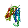
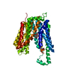
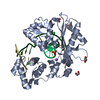
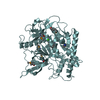
Mass: 26393.756 Da / Num. of mol.: 1
Source method: isolated from a genetically manipulated source
Details: The expressed protein corresponds to residues 71-284 of UniProtKB - G8N1K9. However, sequence alignments indicate that Met46 in this sequence represents the true start and thus the numbering ...Details: The expressed protein corresponds to residues 71-284 of UniProtKB - G8N1K9. However, sequence alignments indicate that Met46 in this sequence represents the true start and thus the numbering used in this PDB entry reflects this i.e Met46 is now Met1. Therefore, in this revised numbering scheme the crystallized protein contains residues 26-239 of the wild type sequence. To this is appended an N-terminal methionine and a C-terminal sequence of KLAAALEHHHHHH, the latter being the nickel affinity tag from the pET21b expression plasmid.
Source: (gene. exp.) Geobacillus thermoleovorans CCB_US3_UF5 (bacteria)
Gene: noc, GTCCBUS3UF5_39100 / Production host: Escherichia coli BL21(DE3) (bacteria) / Strain (production host): BL21(DE3) / Variant (production host): Rosetta / References: UniProt: G8N1K9
#2: Chemical ChemComp-SO4 / SULFATE ION


Mass: 96.063 Da / Num. of mol.: 1 / Source method: obtained synthetically / Formula: SO4
Has ligand of interestN

-
Experimental details

-
Experiment

ExperimentMethod: X-RAY DIFFRACTION / Number of used crystals: 1

-
Sample preparation

CrystalDensity Matthews: 2.24 Å3/Da / Density % sol: 45.2 % / Description: NULL
Crystal growTemperature: 293 K / Method: vapor diffusion, sitting drop / pH: 8 / Details: NULL

-
Data collection

DiffractionMean temperature: 100 K / Serial crystal experiment: N
Diffraction sourceSource: SYNCHROTRON / Site: Diamond / Beamline: I04 / Wavelength: 0.9795 Å
DetectorType: DECTRIS EIGER2 XE 16M / Detector: PIXEL / Date: Aug 11, 2020
RadiationProtocol: SINGLE WAVELENGTH / Monochromatic (M) / Laue (L): M / Scattering type: x-ray
Radiation wavelengthWavelength: 0.9795 Å / Relative weight: 1
ReflectionResolution: 2.95→37.44 Å / Num. obs: 5285 / % possible obs: 100 % / Redundancy: 12.7 % / CC1/2: 0.997 / Rmerge(I) obs: 0.281 / Rpim(I) all: 0.082 / Rrim(I) all: 0.293 / Net I/σ(I): 5.5
Reflection shell

Diffraction-ID: 1

Resolution (Å)Redundancy (%)Rmerge(I) obsNum. unique obsCC1/2Rpim(I) allRrim(I) all% possible all
2.95-3.1312.71.3438350.8550.3881.399100
8.85-37.4410.70.0822910.0250.08499.3

-
Processing

Software
NameVersionClassification
Aimless0.7.4data scaling
REFMAC5.8.0267refinement
PDB_EXTRACT3.27data extraction
DIALSdata reduction
PHASERphasing
RefinementMethod to determine structure: MOLECULAR REPLACEMENT
Starting model: 7NFU
Resolution: 2.95→37.44 Å / Cor.coef. Fo:Fc: 0.923 / Cor.coef. Fo:Fc free: 0.928 / SU B: 31.513 / SU ML: 0.524 / Cross valid method: THROUGHOUT / σ(F): 0 / ESU R Free: 0.498 / Stereochemistry target values: MAXIMUM LIKELIHOOD
Details: HYDROGENS HAVE BEEN ADDED IN THE RIDING POSITIONS U VALUES : REFINED INDIVIDUALLY
RfactorNum. reflection% reflectionSelection details
Rfree0.2881 522 9.9 %RANDOM
Rwork0.2673 ---
obs0.2693 4752 99.89 %-
Solvent computationIon probe radii: 0.8 Å / Shrinkage radii: 0.8 Å / VDW probe radii: 1.2 Å / Solvent model: MASK
Displacement parametersBiso max: 141.43 Å2 / Biso mean: 74.312 Å2 / Biso min: 37.78 Å2
Baniso -1Baniso -2Baniso -3
1-12.86 Å2-0 Å2-0 Å2
2---5.74 Å2-0 Å2
3----7.12 Å2
Refinement stepCycle: final / Resolution: 2.95→37.44 Å
ProteinNucleic acidLigandSolventTotal
Num. atoms1515 0 5 0 1520
Biso mean--69.66 --
Num. residues----199
Refine LS restraints
Refine-IDTypeDev idealDev ideal targetNumber
X-RAY DIFFRACTIONr_bond_refined_d0.0070.0131537
X-RAY DIFFRACTIONr_bond_other_d0.0010.0171511
X-RAY DIFFRACTIONr_angle_refined_deg1.3311.6362090
X-RAY DIFFRACTIONr_angle_other_deg1.3651.5743455
X-RAY DIFFRACTIONr_dihedral_angle_1_deg4.7165198
X-RAY DIFFRACTIONr_dihedral_angle_2_deg38.44123.42176
X-RAY DIFFRACTIONr_dihedral_angle_3_deg18.86915273
X-RAY DIFFRACTIONr_dihedral_angle_4_deg18.9921510
X-RAY DIFFRACTIONr_chiral_restr0.0660.2221
X-RAY DIFFRACTIONr_gen_planes_refined0.0050.021736
X-RAY DIFFRACTIONr_gen_planes_other0.0020.02316
LS refinement shellResolution: 2.95→3.027 Å / Rfactor Rfree error: 0 / Total num. of bins used: 20
RfactorNum. reflection% reflection
Rfree0.443 40 -
Rwork0.438 342 -
all-382 -
obs--100 %

+
About Yorodumi

-
News

-
Feb 9, 2022. New format data for meta-information of EMDB entries

New format data for meta-information of EMDB entries

  • Version 3 of the EMDB header file is now the official format.
  • The previous official version 1.9 will be removed from the archive.

Related info.:EMDB header

External links:wwPDB to switch to version 3 of the EMDB data model

-
Aug 12, 2020. Covid-19 info

Covid-19 info

URL: https://pdbjlvh1.pdbj.org/emnavi/covid19.php

New page: Covid-19 featured information page in EM Navigator.

Related info.:Covid-19 info / Mar 5, 2020. Novel coronavirus structure data

+
Mar 5, 2020. Novel coronavirus structure data

Novel coronavirus structure data

Related info.:Yorodumi Speices / Aug 12, 2020. Covid-19 info

External links:COVID-19 featured content - PDBj / Molecule of the Month (242):Coronavirus Proteases

+
Jan 31, 2019. EMDB accession codes are about to change! (news from PDBe EMDB page)

EMDB accession codes are about to change! (news from PDBe EMDB page)

  • The allocation of 4 digits for EMDB accession codes will soon come to an end. Whilst these codes will remain in use, new EMDB accession codes will include an additional digit and will expand incrementally as the available range of codes is exhausted. The current 4-digit format prefixed with “EMD-” (i.e. EMD-XXXX) will advance to a 5-digit format (i.e. EMD-XXXXX), and so on. It is currently estimated that the 4-digit codes will be depleted around Spring 2019, at which point the 5-digit format will come into force.
  • The EM Navigator/Yorodumi systems omit the EMD- prefix.

Related info.:Q: What is EMD? / ID/Accession-code notation in Yorodumi/EM Navigator

External links:EMDB Accession Codes are Changing Soon! / Contact to PDBj

+
Jul 12, 2017. Major update of PDB

Major update of PDB

  • wwPDB released updated PDB data conforming to the new PDBx/mmCIF dictionary.
  • This is a major update changing the version number from 4 to 5, and with Remediation, in which all the entries are updated.
  • In this update, many items about electron microscopy experimental information are reorganized (e.g. em_software).
  • Now, EM Navigator and Yorodumi are based on the updated data.

External links:wwPDB Remediation / Enriched Model Files Conforming to OneDep Data Standards Now Available in the PDB FTP Archive

-
Yorodumi

Thousand views of thousand structures

  • Yorodumi is a browser for structure data from EMDB, PDB, SASBDB, etc.
  • This page is also the successor to EM Navigator detail page, and also detail information page/front-end page for Omokage search.
  • The word "yorodu" (or yorozu) is an old Japanese word meaning "ten thousand". "mi" (miru) is to see.

Related info.:EMDB / PDB / SASBDB / Comparison of 3 databanks / Yorodumi Search / Aug 31, 2016. New EM Navigator & Yorodumi / Yorodumi Papers / Jmol/JSmol / Function and homology information / Changes in new EM Navigator and Yorodumi

Read more