 Movie
Movie Controller

[English] 日本語
 Yorodumi
- PDB-7nb5: Structure of EstD11 S144A in complex with naproxen p-nitrophenol ester -
+ Open data

- Basic information
| Entry | Database: PDB / ID: 7nb5 | |||||||||
|---|---|---|---|---|---|---|---|---|---|---|
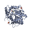
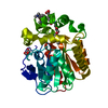
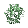
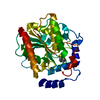
| Title | Structure of EstD11 S144A in complex with naproxen p-nitrophenol ester | |||||||||
 Components Components | EstD11 S144A | |||||||||
 Keywords Keywords | HYDROLASE / Esterase Hormone-Sensitive Lipase Metagenome library Crystal structure | |||||||||
| Function / homology | Alpha/Beta hydrolase fold, catalytic domain / Rossmann fold / 3-Layer(aba) Sandwich / Alpha Beta / Chem-U68 Function and homology information Function and homology information | |||||||||
| Biological species |  uncultured bacterium (environmental samples) uncultured bacterium (environmental samples) | |||||||||
| Method |  X-RAY DIFFRACTION / SYNCHROTRON / MOLECULAR REPLACEMENT / Resolution: 2.13 Å X-RAY DIFFRACTION / SYNCHROTRON / MOLECULAR REPLACEMENT / Resolution: 2.13 Å | |||||||||
 Authors Authors | Miguel-Ruano, V. / Rivera, I. / Hermoso, J.A. | |||||||||
| Funding support |  Spain, 1items Spain, 1items
| |||||||||
 Citation Citation | Journal: Comput Struct Biotechnol J / Year: 2021 Title: Biochemical and Structural Characterization of a novel thermophilic esterase EstD11 provide catalytic insights for the HSL family. Authors: Miguel-Ruano, V. / Rivera, I. / Rajkovic, J. / Knapik, K. / Torrado, A. / Otero, J.M. / Beneventi, E. / Becerra, M. / Sanchez-Costa, M. / Hidalgo, A. / Berenguer, J. / Gonzalez-Siso, M.I. / ...Authors: Miguel-Ruano, V. / Rivera, I. / Rajkovic, J. / Knapik, K. / Torrado, A. / Otero, J.M. / Beneventi, E. / Becerra, M. / Sanchez-Costa, M. / Hidalgo, A. / Berenguer, J. / Gonzalez-Siso, M.I. / Cruces, J. / Rua, M.L. / Hermoso, J.A. | |||||||||
| History |
|
- Structure visualization
| Structure viewer | Molecule: MolmilJmol/JSmol |
|---|
- Downloads & links
-Download
| PDBx/mmCIF format | 7nb5.cif.gz | 130.6 KB | Display | PDBx/mmCIF format |
|---|---|---|---|---|
| PDB format | pdb7nb5.ent.gz | 100.5 KB | Display | PDB format |
| PDBx/mmJSON format | 7nb5.json.gz | Tree view | PDBx/mmJSON format | |
| Others |  Other downloads Other downloads |
-Validation report
| Arichive directory | https://data.pdbj.org/pub/pdb/validation_reports/nb/7nb5ftp://data.pdbj.org/pub/pdb/validation_reports/nb/7nb5 | HTTPS FTP |
|---|
-Related structure data
| Related structure data | 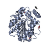 7at0SC 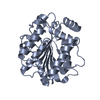 7at2C  7at3C  7at4C 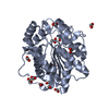 7atdC  7atfC  7atqC 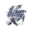 7auyC  7av5C S: Starting model for refinement C: citing same article ( |
|---|---|
| Similar structure data |


-Links
 PDBj
PDBj- Assembly
| Deposited unit | 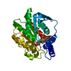
| ||||||||||||
|---|---|---|---|---|---|---|---|---|---|---|---|---|---|
| 1 |
| ||||||||||||
| Unit cell |
| ||||||||||||
| Components on special symmetry positions |
|
-Components
| #1: Protein | Mass: 32038.812 Da / Num. of mol.: 1 Source method: isolated from a genetically manipulated source Source: (gene. exp.) uncultured bacterium (environmental samples)Production host: |
|---|---|
| #2: Chemical | ChemComp-U68 /   Mass: 351.353 Da / Num. of mol.: 1 / Source method: obtained synthetically / Formula: C20H17NO5 / Feature type: SUBJECT OF INVESTIGATION Mass: 351.353 Da / Num. of mol.: 1 / Source method: obtained synthetically / Formula: C20H17NO5 / Feature type: SUBJECT OF INVESTIGATION |
| #3: Water | ChemComp-HOH /  Mass: 18.015 Da / Num. of mol.: 99 / Source method: isolated from a natural source / Formula: H2O Mass: 18.015 Da / Num. of mol.: 99 / Source method: isolated from a natural source / Formula: H2O |
| Has ligand of interest | Y |
| Has protein modification | N |
-Experimental details
-Experiment
| Experiment | Method: X-RAY DIFFRACTION / Number of used crystals: 1 |
|---|
- Sample preparation
| Crystal | Density Matthews: 2.11 Å3/Da / Density % sol: 41.78 % |
|---|---|
| Crystal grow | Temperature: 291 K / Method: vapor diffusion, sitting drop / pH: 5 / Details: Sodium formate 3.2M and citrate pH 5 0.1M |
-Data collection
| Diffraction | Mean temperature: 100 K / Serial crystal experiment: N |
|---|---|
| Diffraction source | Source: SYNCHROTRON / Site: ALBA / Beamline: XALOC / Wavelength: 0.979 Å |
| Detector | Type: DECTRIS PILATUS 6M / Detector: PIXEL / Date: Nov 20, 2020 |
| Radiation | Protocol: SINGLE WAVELENGTH / Monochromatic (M) / Laue (L): M / Scattering type: x-ray |
| Radiation wavelength | Wavelength: 0.979 Å / Relative weight: 1 |
| Reflection | Resolution: 2.13→44.84 Å / Num. obs: 15581 / % possible obs: 99.9 % / Redundancy: 6.7 % / CC1/2: 0.998 / Rmerge(I) obs: 0.067 / Rpim(I) all: 0.027 / Net I/σ(I): 18.2 |
| Reflection shell | Resolution: 2.13→2.19 Å / Rmerge(I) obs: 0.231 / Mean I/σ(I) obs: 8.2 / Num. unique obs: 1265 / CC1/2: 0.989 / Rpim(I) all: 0.09 / % possible all: 100 |
- Processing
| Software |
| |||||||||||||||||||||||||||||||||||||||||||||||||||||||||||||||||||||||||||||||||||||||||||||||||||||||||
|---|---|---|---|---|---|---|---|---|---|---|---|---|---|---|---|---|---|---|---|---|---|---|---|---|---|---|---|---|---|---|---|---|---|---|---|---|---|---|---|---|---|---|---|---|---|---|---|---|---|---|---|---|---|---|---|---|---|---|---|---|---|---|---|---|---|---|---|---|---|---|---|---|---|---|---|---|---|---|---|---|---|---|---|---|---|---|---|---|---|---|---|---|---|---|---|---|---|---|---|---|---|---|---|---|---|---|
| Refinement | Method to determine structure: MOLECULAR REPLACEMENT Starting model: 7AT0 Resolution: 2.13→42.36 Å / Cor.coef. Fo:Fc: 0.963 / Cor.coef. Fo:Fc free: 0.931 / SU B: 11.629 / SU ML: 0.153 / Cross valid method: THROUGHOUT / ESU R: 0.244 / ESU R Free: 0.198 / Stereochemistry target values: MAXIMUM LIKELIHOOD Details: U VALUES : WITH TLS ADDED HYDROGENS HAVE BEEN ADDED IN THE RIDING POSITIONS U VALUES : RESIDUAL ONLY
| |||||||||||||||||||||||||||||||||||||||||||||||||||||||||||||||||||||||||||||||||||||||||||||||||||||||||
| Solvent computation | Ion probe radii: 0.8 Å / Shrinkage radii: 0.8 Å / VDW probe radii: 1.1 Å / Solvent model: MASK | |||||||||||||||||||||||||||||||||||||||||||||||||||||||||||||||||||||||||||||||||||||||||||||||||||||||||
| Displacement parameters | Biso mean: 39.253 Å2
| |||||||||||||||||||||||||||||||||||||||||||||||||||||||||||||||||||||||||||||||||||||||||||||||||||||||||
| Refinement step | Cycle: LAST / Resolution: 2.13→42.36 Å
| |||||||||||||||||||||||||||||||||||||||||||||||||||||||||||||||||||||||||||||||||||||||||||||||||||||||||
| Refine LS restraints |
| |||||||||||||||||||||||||||||||||||||||||||||||||||||||||||||||||||||||||||||||||||||||||||||||||||||||||
| LS refinement shell | Resolution: 2.13→2.185 Å / Total num. of bins used: 20
| |||||||||||||||||||||||||||||||||||||||||||||||||||||||||||||||||||||||||||||||||||||||||||||||||||||||||
| Refinement TLS params. | Method: refined / Origin x: 13.576 Å / Origin y: 19.592 Å / Origin z: 16.272 Å
|
