 Movie
Movie Controller


+ Open data

- Basic information
| Entry | Database: PDB / ID: 7n9z | |||||||||||||||||||||||||||||||||||||||
|---|---|---|---|---|---|---|---|---|---|---|---|---|---|---|---|---|---|---|---|---|---|---|---|---|---|---|---|---|---|---|---|---|---|---|---|---|---|---|---|---|
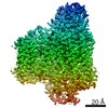
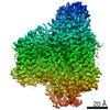
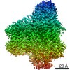
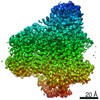
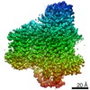
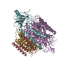
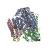
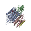
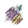
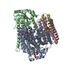
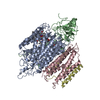
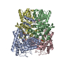
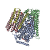
| Title | E. coli cytochrome bo3 in MSP nanodisc | |||||||||||||||||||||||||||||||||||||||
 Components Components |
| |||||||||||||||||||||||||||||||||||||||
 Keywords Keywords | MEMBRANE PROTEIN / Ubiquinone / Heme-copper Oxidoreductase / Electron transport / Bioenergetics / Proton pump | |||||||||||||||||||||||||||||||||||||||
| Function / homology |  Function and homology information Function and homology informationcytochrome bo3 ubiquinol oxidase activity => GO:0009486 / ubiquinol oxidase (H+-transporting) / cytochrome bo3 ubiquinol oxidase activity / : / aerobic electron transport chain / oxidoreductase activity, acting on diphenols and related substances as donors, oxygen as acceptor / cytochrome-c oxidase activity / electron transport coupled proton transport / membrane => GO:0016020 / ATP synthesis coupled electron transport ...cytochrome bo3 ubiquinol oxidase activity => GO:0009486 / ubiquinol oxidase (H+-transporting) / cytochrome bo3 ubiquinol oxidase activity / : / aerobic electron transport chain / oxidoreductase activity, acting on diphenols and related substances as donors, oxygen as acceptor / cytochrome-c oxidase activity / electron transport coupled proton transport / membrane => GO:0016020 / ATP synthesis coupled electron transport / aerobic respiration / copper ion binding / heme binding / plasma membrane Similarity search - Function | |||||||||||||||||||||||||||||||||||||||
| Biological species |  | |||||||||||||||||||||||||||||||||||||||
| Method | ELECTRON MICROSCOPY / single particle reconstruction / cryo EM / Resolution: 2.19 Å | |||||||||||||||||||||||||||||||||||||||
 Authors Authors | Vallese, F. / Clarke, O.B. | |||||||||||||||||||||||||||||||||||||||
 Citation Citation | Journal: Proc Natl Acad Sci U S A / Year: 2021 Title: Cryo-EM structures of cytochrome reveal bound phospholipids and ubiquinone-8 in a dynamic substrate binding site. Authors: Jiao Li / Long Han / Francesca Vallese / Ziqiao Ding / Sylvia K Choi / Sangjin Hong / Yanmei Luo / Bin Liu / Chun Kit Chan / Emad Tajkhorshid / Jiapeng Zhu / Oliver Clarke / Kai Zhang / Robert Gennis /   Abstract: Two independent structures of the proton-pumping, respiratory cytochrome ubiquinol oxidase (cyt ) have been determined by cryogenic electron microscopy (cryo-EM) in styrene-maleic acid (SMA) ...Two independent structures of the proton-pumping, respiratory cytochrome ubiquinol oxidase (cyt ) have been determined by cryogenic electron microscopy (cryo-EM) in styrene-maleic acid (SMA) copolymer nanodiscs and in membrane scaffold protein (MSP) nanodiscs to 2.55- and 2.19-Å resolution, respectively. The structures include the metal redox centers (heme , heme , and Cu), the redox-active cross-linked histidine-tyrosine cofactor, and the internal water molecules in the proton-conducting D channel. Each structure also contains one equivalent of ubiquinone-8 (UQ8) in the substrate binding site as well as several phospholipid molecules. The isoprene side chain of UQ8 is clamped within a hydrophobic groove in subunit I by transmembrane helix TM0, which is only present in quinol oxidases and not in the closely related cytochrome oxidases. Both structures show carbonyl O1 of the UQ8 headgroup hydrogen bonded to D75 and R71 In both structures, residue H98 occupies two conformations. In conformation 1, H98 forms a hydrogen bond with carbonyl O4 of the UQ8 headgroup, but in conformation 2, the imidazole side chain of H98 has flipped to form a hydrogen bond with E14 at the N-terminal end of TM0. We propose that H98 dynamics facilitate proton transfer from ubiquinol to the periplasmic aqueous phase during oxidation of the substrate. Computational studies show that TM0 creates a channel, allowing access of water to the ubiquinol headgroup and to H98. | |||||||||||||||||||||||||||||||||||||||
| History |
|
- Structure visualization
| Movie |
Movie viewer |
|---|---|
| Structure viewer | Molecule: MolmilJmol/JSmol |
- Downloads & links
-Download
| PDBx/mmCIF format | 7n9z.cif.gz | 449.7 KB | Display | PDBx/mmCIF format |
|---|---|---|---|---|
| PDB format | pdb7n9z.ent.gz | 369.5 KB | Display | PDB format |
| PDBx/mmJSON format | 7n9z.json.gz | Tree view | PDBx/mmJSON format | |
| Others |  Other downloads Other downloads |
-Validation report
| Arichive directory | https://data.pdbj.org/pub/pdb/validation_reports/n9/7n9zftp://data.pdbj.org/pub/pdb/validation_reports/n9/7n9z | HTTPS FTP |
|---|
-Related structure data
| Related structure data | 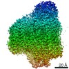 24265MC 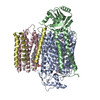 7cubC 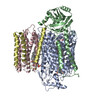 7cuqC  7cuwC C: citing same article ( M: map data used to model this data |
|---|---|
| Similar structure data | |
| EM raw data | EMPIAR-10737 (Title: Cryo-EM structures of E. coli cytochrome bo3 in MSP Nanodiscs Data size: 851.4 Data #1: Unaligned multi-frame micrographs of cyt bo3 in MSP Nanodiscs [micrographs - multiframe]) |

-Links
 PDBj
PDBj


- Assembly
| Deposited unit | 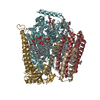
|
|---|---|
| 1 |
|
-Components
-Cytochrome o ubiquinol oxidase, subunit ... , 2 types, 2 molecules FI
| #1: Protein | Mass: 74424.469 Da / Num. of mol.: 1 / Source method: isolated from a natural source / Source: (natural) References: UniProt: H4KCU1, Oxidoreductases; Acting on diphenols and related substances as donors; With oxygen as acceptor |
|---|---|
| #4: Protein | Mass: 12037.402 Da / Num. of mol.: 1 / Source method: isolated from a natural source / Source: (natural) References: UniProt: I2RK84, Oxidoreductases; Acting on diphenols and related substances as donors; With oxygen as acceptor |
-Protein , 2 types, 2 molecules GH
| #2: Protein | Mass: 34947.203 Da / Num. of mol.: 1 / Source method: isolated from a natural source / Source: (natural) |
|---|---|
| #3: Protein | Mass: 22642.566 Da / Num. of mol.: 1 / Source method: isolated from a natural source / Source: (natural) |
-Non-polymers , 9 types, 267 molecules 


| #5: Chemical | ChemComp-UQ8 /  Mass: 727.109 Da / Num. of mol.: 1 / Source method: obtained synthetically / Formula: C49H74O4 / Feature type: SUBJECT OF INVESTIGATION Mass: 727.109 Da / Num. of mol.: 1 / Source method: obtained synthetically / Formula: C49H74O4 / Feature type: SUBJECT OF INVESTIGATION | ||||||||||||||
|---|---|---|---|---|---|---|---|---|---|---|---|---|---|---|---|
| #6: Chemical | ChemComp-LHG /  Mass: 722.970 Da / Num. of mol.: 9 / Source method: obtained synthetically / Formula: C38H75O10P / Feature type: SUBJECT OF INVESTIGATION / Comment: phospholipid*YM Mass: 722.970 Da / Num. of mol.: 9 / Source method: obtained synthetically / Formula: C38H75O10P / Feature type: SUBJECT OF INVESTIGATION / Comment: phospholipid*YM#7: Chemical |  Mass: 748.065 Da / Num. of mol.: 2 / Source method: obtained synthetically / Formula: C41H82NO8P / Feature type: SUBJECT OF INVESTIGATION / Comment: phospholipid*YM Mass: 748.065 Da / Num. of mol.: 2 / Source method: obtained synthetically / Formula: C41H82NO8P / Feature type: SUBJECT OF INVESTIGATION / Comment: phospholipid*YM#8: Chemical | ChemComp-HEO / |  Mass: 838.854 Da / Num. of mol.: 1 / Source method: obtained synthetically / Formula: C49H58FeN4O5 / Feature type: SUBJECT OF INVESTIGATION Mass: 838.854 Da / Num. of mol.: 1 / Source method: obtained synthetically / Formula: C49H58FeN4O5 / Feature type: SUBJECT OF INVESTIGATION#9: Chemical | ChemComp-HEM / |  Mass: 616.487 Da / Num. of mol.: 1 / Source method: obtained synthetically / Formula: C34H32FeN4O4 / Feature type: SUBJECT OF INVESTIGATION Mass: 616.487 Da / Num. of mol.: 1 / Source method: obtained synthetically / Formula: C34H32FeN4O4 / Feature type: SUBJECT OF INVESTIGATION#10: Chemical | ChemComp-CDL / |  Mass: 1464.043 Da / Num. of mol.: 1 / Source method: obtained synthetically / Formula: C81H156O17P2 / Feature type: SUBJECT OF INVESTIGATION / Comment: phospholipid*YM Mass: 1464.043 Da / Num. of mol.: 1 / Source method: obtained synthetically / Formula: C81H156O17P2 / Feature type: SUBJECT OF INVESTIGATION / Comment: phospholipid*YM#11: Chemical | ChemComp-CU / |  Mass: 63.546 Da / Num. of mol.: 1 / Source method: obtained synthetically / Formula: Cu / Feature type: SUBJECT OF INVESTIGATION Mass: 63.546 Da / Num. of mol.: 1 / Source method: obtained synthetically / Formula: Cu / Feature type: SUBJECT OF INVESTIGATION#12: Chemical | ChemComp-ZN / |  Mass: 65.409 Da / Num. of mol.: 1 / Source method: obtained synthetically / Formula: Zn / Feature type: SUBJECT OF INVESTIGATION Mass: 65.409 Da / Num. of mol.: 1 / Source method: obtained synthetically / Formula: Zn / Feature type: SUBJECT OF INVESTIGATION#13: Water | ChemComp-HOH / | Mass: 18.015 Da / Num. of mol.: 250 / Source method: isolated from a natural source / Formula: H2O |
-Details
| Has ligand of interest | Y |
|---|---|
| Has protein modification | N |
-Experimental details
-Experiment
| Experiment | Method: ELECTRON MICROSCOPY |
|---|---|
| EM experiment | Aggregation state: PARTICLE / 3D reconstruction method: single particle reconstruction |
- Sample preparation
| Component | Name: Cytochrome bo 3 ubiquinol oxidase / Type: COMPLEX / Entity ID: #1-#4 / Source: NATURAL | ||||||||||||
|---|---|---|---|---|---|---|---|---|---|---|---|---|---|
| Molecular weight | Experimental value: NO | ||||||||||||
| Source (natural) | Organism: | ||||||||||||
| Buffer solution | pH: 7.4 | ||||||||||||
| Buffer component |
| ||||||||||||
| Specimen | Conc.: 2.6 mg/ml / Embedding applied: NO / Shadowing applied: NO / Staining applied: NO / Vitrification applied: YES | ||||||||||||
| Specimen support | Grid material: GOLD / Grid mesh size: 300 divisions/in. / Grid type: UltrAuFoil R0.6/1 | ||||||||||||
| Vitrification | Instrument: FEI VITROBOT MARK IV / Cryogen name: ETHANE / Humidity: 100 % / Chamber temperature: 277 K |
- Electron microscopy imaging
| Experimental equipment |  Model: Titan Krios / Image courtesy: FEI Company |
|---|---|
| Microscopy | Model: FEI TITAN KRIOS |
| Electron gun | Electron source:  FIELD EMISSION GUN / Accelerating voltage: 300 kV / Illumination mode: FLOOD BEAM FIELD EMISSION GUN / Accelerating voltage: 300 kV / Illumination mode: FLOOD BEAM |
| Electron lens | Mode: BRIGHT FIELD |
| Image recording | Electron dose: 58 e/Å2 / Film or detector model: GATAN K3 (6k x 4k) |
- Processing
| Software | Name: PHENIX / Version: 1.19.2_4158: / Classification: refinement | ||||||||||||||||||||||||
|---|---|---|---|---|---|---|---|---|---|---|---|---|---|---|---|---|---|---|---|---|---|---|---|---|---|
| EM software | Name: PHENIX / Category: model refinement | ||||||||||||||||||||||||
| CTF correction | Type: PHASE FLIPPING AND AMPLITUDE CORRECTION | ||||||||||||||||||||||||
| 3D reconstruction | Resolution: 2.19 Å / Resolution method: FSC 0.143 CUT-OFF / Num. of particles: 94681 / Symmetry type: POINT | ||||||||||||||||||||||||
| Atomic model building | PDB-ID: 1FFT Accession code: 1FFT / Source name: PDB / Type: experimental model | ||||||||||||||||||||||||
| Refinement | Highest resolution: 2.19 Å | ||||||||||||||||||||||||
| Refine LS restraints |
|

