 Movie
Movie Controller

[English] 日本語
 Yorodumi
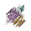
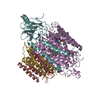
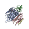
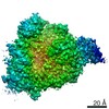
- PDB-6koc: X-ray Structure of the proton-pumping cytochrome aa3-600 menaquin... -
+ Open data

- Basic information
| Entry | Database: PDB / ID: 6koc | ||||||
|---|---|---|---|---|---|---|---|
| Title | X-ray Structure of the proton-pumping cytochrome aa3-600 menaquinol oxidase from Bacillus subtilis complexed with 3-iodo-N-oxo-2-heptyl-4-hydroxyquinoline | ||||||
 Components Components |
| ||||||
 Keywords Keywords | OXIDOREDUCTASE / Menaquinol oxidase / Complex / Proton pumping / inhibitor | ||||||
| Function / homology |  Function and homology information Function and homology informationOxidoreductases; Acting on diphenols and related substances as donors; With oxygen as acceptor / cytochrome o ubiquinol oxidase complex / cytochrome bo3 ubiquinol oxidase activity / aerobic electron transport chain / respiratory chain complex / oxidoreductase activity, acting on diphenols and related substances as donors, oxygen as acceptor / oxidative phosphorylation / cytochrome-c oxidase activity / proton transmembrane transporter activity / electron transport coupled proton transport ...Oxidoreductases; Acting on diphenols and related substances as donors; With oxygen as acceptor / cytochrome o ubiquinol oxidase complex / cytochrome bo3 ubiquinol oxidase activity / aerobic electron transport chain / respiratory chain complex / oxidoreductase activity, acting on diphenols and related substances as donors, oxygen as acceptor / oxidative phosphorylation / cytochrome-c oxidase activity / proton transmembrane transporter activity / electron transport coupled proton transport / ATP synthesis coupled electron transport / aerobic respiration / respiratory electron transport chain / oxidoreductase activity / membrane raft / copper ion binding / heme binding / plasma membrane Similarity search - Function | ||||||
| Biological species |  | ||||||
| Method |  X-RAY DIFFRACTION / SYNCHROTRON / MOLECULAR REPLACEMENT / Resolution: 3.8 Å X-RAY DIFFRACTION / SYNCHROTRON / MOLECULAR REPLACEMENT / Resolution: 3.8 Å | ||||||
 Authors Authors | Xu, J. / Ding, Z. / Liu, B. / Li, J. / Gennis, R.B. / Zhu, J. | ||||||
 Citation Citation | Journal: Proc.Natl.Acad.Sci.USA / Year: 2020 Title: Structure of the cytochromeaa3-600 heme-copper menaquinol oxidase bound to inhibitor HQNO shows TM0 is part of the quinol binding site. Authors: Xu, J. / Ding, Z. / Liu, B. / Yi, S.M. / Li, J. / Zhang, Z. / Liu, Y. / Li, J. / Liu, L. / Zhou, A. / Gennis, R.B. / Zhu, J. | ||||||
| History |
|
- Structure visualization
| Structure viewer | Molecule: MolmilJmol/JSmol |
|---|
- Downloads & links
-Download
| PDBx/mmCIF format | 6koc.cif.gz | 975.5 KB | Display | PDBx/mmCIF format |
|---|---|---|---|---|
| PDB format | pdb6koc.ent.gz | 775.5 KB | Display | PDB format |
| PDBx/mmJSON format | 6koc.json.gz | Tree view | PDBx/mmJSON format | |
| Others |  Other downloads Other downloads |
-Validation report
| Arichive directory | https://data.pdbj.org/pub/pdb/validation_reports/ko/6kocftp://data.pdbj.org/pub/pdb/validation_reports/ko/6koc | HTTPS FTP |
|---|
-Related structure data
| Related structure data | 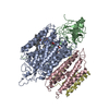 6kobSC  6koeC S: Starting model for refinement C: citing same article ( |
|---|---|
| Similar structure data |


-Links
 PDBj
PDBj


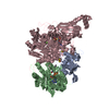
- Assembly
| Deposited unit | 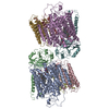
| ||||||||||
|---|---|---|---|---|---|---|---|---|---|---|---|
| 1 | 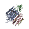
| ||||||||||
| 2 | 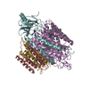
| ||||||||||
| Unit cell |
|
-Components
-AA3-600 quinol oxidase subunit ... , 2 types, 4 molecules AECG
| #1: Protein | Mass: 73892.789 Da / Num. of mol.: 2 Source method: isolated from a genetically manipulated source Source: (gene. exp.) Gene: qoxB, B4122_4931, B4417_2140, ETA10_20065, ETK61_21170, ETL41_11350, SC09_contig4orf01211 Production host: #3: Protein | Mass: 22689.572 Da / Num. of mol.: 2 Source method: isolated from a genetically manipulated source Source: (gene. exp.) Gene: B4122_4930, B4417_2139, ETA10_20060, ETK61_21165, ETL41_11345, SC09_contig4orf01209 Production host: |
|---|
-Quinol oxidase subunit ... , 2 types, 4 molecules BFDH
| #2: Protein | Mass: 33589.961 Da / Num. of mol.: 2 Source method: isolated from a genetically manipulated source Source: (gene. exp.) References: UniProt: A0A2I7T8S1, UniProt: P34957*PLUS, Oxidoreductases; Acting on diphenols and related substances as donors; With oxygen as acceptor #4: Protein | Mass: 12319.210 Da / Num. of mol.: 2 Source method: isolated from a genetically manipulated source Source: (gene. exp.) Strain: 168 / Gene: qoxD, BSU38140, ipa-40d / Production host: References: UniProt: P34959, Oxidoreductases; Acting on diphenols and related substances as donors; With oxygen as acceptor |
|---|
-Non-polymers , 3 types, 8 molecules 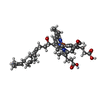

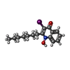
| #5: Chemical | ChemComp-HEA /  Mass: 852.837 Da / Num. of mol.: 4 / Source method: obtained synthetically / Formula: C49H56FeN4O6 Mass: 852.837 Da / Num. of mol.: 4 / Source method: obtained synthetically / Formula: C49H56FeN4O6#6: Chemical |  Mass: 63.546 Da / Num. of mol.: 2 / Source method: obtained synthetically / Formula: Cu Mass: 63.546 Da / Num. of mol.: 2 / Source method: obtained synthetically / Formula: Cu#7: Chemical |  Mass: 385.240 Da / Num. of mol.: 2 / Source method: obtained synthetically / Formula: C16H20INO2 Mass: 385.240 Da / Num. of mol.: 2 / Source method: obtained synthetically / Formula: C16H20INO2 |
|---|
-Details
| Has ligand of interest | N |
|---|---|
| Sequence details | Authors know the sequence of chain D/H, but they are not sure of the alignment for first 22 ...Authors know the sequence of chain D/H, but they are not sure of the alignment for first 22 residues in the coordinates. The residue numbers 0-21 in the coordinates may be meaningless. The correct sequence is ANKSAEHSHF |
-Experimental details
-Experiment
| Experiment | Method: X-RAY DIFFRACTION / Number of used crystals: 1 |
|---|
- Sample preparation
| Crystal | Density Matthews: 4.62 Å3/Da / Density % sol: 76.6 % |
|---|---|
| Crystal grow | Temperature: 295 K / Method: vapor diffusion, sitting drop / pH: 6.3 Details: 0.1 M Calcium chloride, 0.1M Tris pH 6.3, 13% PEG 2000 MME |
-Data collection
| Diffraction | Mean temperature: 100 K / Serial crystal experiment: N |
|---|---|
| Diffraction source | Source: SYNCHROTRON / Site: SSRF  / Beamline: BL17U1 / Wavelength: 0.9793 Å / Beamline: BL17U1 / Wavelength: 0.9793 Å |
| Detector | Type: DECTRIS PILATUS3 6M / Detector: PIXEL / Date: Nov 26, 2018 |
| Radiation | Protocol: SINGLE WAVELENGTH / Monochromatic (M) / Laue (L): M / Scattering type: x-ray |
| Radiation wavelength | Wavelength: 0.9793 Å / Relative weight: 1 |
| Reflection | Resolution: 3.8→48.28 Å / Num. obs: 50255 / % possible obs: 98.5 % / Redundancy: 2.6 % / CC1/2: 0.878 / Rmerge(I) obs: 0.22 / Net I/σ(I): 3.4 |
| Reflection shell | Resolution: 3.8→3.92 Å / Rmerge(I) obs: 1.264 / Num. unique obs: 4597 / CC1/2: 0.382 |
- Processing
| Software |
| ||||||||||||||||||||||||||||||||||||||||
|---|---|---|---|---|---|---|---|---|---|---|---|---|---|---|---|---|---|---|---|---|---|---|---|---|---|---|---|---|---|---|---|---|---|---|---|---|---|---|---|---|---|
| Refinement | Method to determine structure: MOLECULAR REPLACEMENT Starting model: 6KOB Resolution: 3.8→48.27 Å / Cross valid method: FREE R-VALUE
| ||||||||||||||||||||||||||||||||||||||||
| Refinement step | Cycle: LAST / Resolution: 3.8→48.27 Å
| ||||||||||||||||||||||||||||||||||||||||
| LS refinement shell | Resolution: 3.8001→3.8951 Å
| ||||||||||||||||||||||||||||||||||||||||
| Refinement TLS params. | Method: refined / Origin x: -24.7077 Å / Origin y: 5.1896 Å / Origin z: -12.9278 Å
| ||||||||||||||||||||||||||||||||||||||||
| Refinement TLS group | Selection details: all |
