[English] 日本語
Yorodumi
- PDB-7n9g: Crystal structure of the Abl 1b Kinase domain in complex with Das... -

+
Open data


ID or keywords:

Loading...

-
Basic information

Entry
Database: PDB / ID: 7n9g
TitleCrystal structure of the Abl 1b Kinase domain in complex with Dasatinib and Imatinib
ComponentsTyrosine-protein kinase ABL1
KeywordsCELL CYCLE / Abl kinase / kinase allosteric regulation / drug resistance
Function / homology
Function and homology information


positive regulation of actin filament binding / negative regulation of ubiquitin-protein transferase activity / positive regulation of oxidoreductase activity / protein localization to cytoplasmic microtubule plus-end / DN4 thymocyte differentiation / response to epinephrine / activation of protein kinase C activity / phospholipase C-inhibiting G protein-coupled receptor signaling pathway / podocyte apoptotic process / delta-catenin binding ...positive regulation of actin filament binding / negative regulation of ubiquitin-protein transferase activity / positive regulation of oxidoreductase activity / protein localization to cytoplasmic microtubule plus-end / DN4 thymocyte differentiation / response to epinephrine / activation of protein kinase C activity / phospholipase C-inhibiting G protein-coupled receptor signaling pathway / podocyte apoptotic process / delta-catenin binding / Role of ABL in ROBO-SLIT signaling / transitional one stage B cell differentiation / regulation of postsynaptic specialization assembly / regulation of modification of synaptic structure / nicotinate-nucleotide adenylyltransferase activity / DNA conformation change / neuroepithelial cell differentiation / : / B cell proliferation involved in immune response / cerebellum morphogenesis / positive regulation of Wnt signaling pathway, planar cell polarity pathway / positive regulation of extracellular matrix organization / microspike assembly / B-1 B cell homeostasis / neuropilin signaling pathway / neuropilin binding / bubble DNA binding / mitochondrial depolarization / regulation of cell motility / activated T cell proliferation / positive regulation of establishment of T cell polarity / cellular response to dopamine / positive regulation of blood vessel branching / proline-rich region binding / regulation of Cdc42 protein signal transduction / syntaxin binding / mitogen-activated protein kinase binding / regulation of hematopoietic stem cell differentiation / myoblast proliferation / alpha-beta T cell differentiation / positive regulation of dendrite development / regulation of axon extension / regulation of T cell differentiation / cardiac muscle cell proliferation / positive regulation of cell migration involved in sprouting angiogenesis / positive regulation of peptidyl-tyrosine phosphorylation / HDR through Single Strand Annealing (SSA) / negative regulation of cell-cell adhesion / Myogenesis / platelet-derived growth factor receptor-beta signaling pathway / positive regulation of osteoblast proliferation / RUNX2 regulates osteoblast differentiation / regulation of microtubule polymerization / Fc-gamma receptor signaling pathway involved in phagocytosis / associative learning / vascular endothelial cell response to oscillatory fluid shear stress / Bergmann glial cell differentiation / regulation of endocytosis / neuromuscular process controlling balance / negative regulation of long-term synaptic potentiation / negative regulation of mitotic cell cycle / negative regulation of cellular senescence / negative regulation of BMP signaling pathway / actin monomer binding / canonical NF-kappaB signal transduction / signal transduction in response to DNA damage / positive regulation of focal adhesion assembly / RHO GTPases Activate WASPs and WAVEs / BMP signaling pathway / endothelial cell migration / positive regulation of T cell migration / negative regulation of double-strand break repair via homologous recombination / mismatch repair / negative regulation of endothelial cell apoptotic process / regulation of cell adhesion / peptidyl-tyrosine autophosphorylation / four-way junction DNA binding / cellular response to transforming growth factor beta stimulus / spleen development / positive regulation of stress fiber assembly / ruffle / positive regulation of vasoconstriction / ephrin receptor binding / actin filament polymerization / positive regulation of substrate adhesion-dependent cell spreading / phosphotyrosine residue binding / positive regulation of endothelial cell migration / ERK1 and ERK2 cascade / positive regulation of interleukin-2 production / positive regulation of mitotic cell cycle / substrate adhesion-dependent cell spreading / SH2 domain binding / response to endoplasmic reticulum stress / positive regulation of release of sequestered calcium ion into cytosol / thymus development / protein kinase C binding / Turbulent (oscillatory, disturbed) flow shear stress activates signaling by PIEZO1 and integrins in endothelial cells / post-embryonic development / integrin-mediated signaling pathway / establishment of localization in cell
Similarity search - Function
F-actin binding / F-actin binding / F-actin binding domain (FABD) / Tyrosine-protein kinase ABL, SH2 domain / : / SH3 domain / SH2 domain / Src homology 2 (SH2) domain profile. / Src homology 2 domains / SH2 domain ...F-actin binding / F-actin binding / F-actin binding domain (FABD) / Tyrosine-protein kinase ABL, SH2 domain / : / SH3 domain / SH2 domain / Src homology 2 (SH2) domain profile. / Src homology 2 domains / SH2 domain / SH2 domain superfamily / Src homology 3 domains / SH3-like domain superfamily / Src homology 3 (SH3) domain profile. / SH3 domain / Tyrosine-protein kinase, catalytic domain / Tyrosine kinase, catalytic domain / Tyrosine protein kinases specific active-site signature. / Tyrosine-protein kinase, active site / Serine-threonine/tyrosine-protein kinase, catalytic domain / Protein tyrosine and serine/threonine kinase / Phosphorylase Kinase; domain 1 / Phosphorylase Kinase; domain 1 / Transferase(Phosphotransferase) domain 1 / Transferase(Phosphotransferase); domain 1 / Protein kinase, ATP binding site / Protein kinases ATP-binding region signature. / Protein kinase domain profile. / Protein kinase domain / Protein kinase-like domain superfamily / 2-Layer Sandwich / Orthogonal Bundle / Mainly Alpha / Alpha Beta
Similarity search - Domain/homology
Chem-1N1 / PHOSPHATE ION / Chem-STI / Tyrosine-protein kinase ABL1
Similarity search - Component
Biological speciesHomo sapiens (human)
MethodX-RAY DIFFRACTION / SYNCHROTRON / MOLECULAR REPLACEMENT / Resolution: 2.2 Å
AuthorsMiller, D.J. / Xie, T.
Funding support United States, 1items
OrganizationGrant numberCountry
National Institutes of Health/Office of the Director5R35GM122462-05 United States
CitationJournal: J.Mol.Biol. / Year: 2022
Title: Imatinib can act as an Allosteric Activator of Abl Kinase.
Authors: Xie, T. / Saleh, T. / Rossi, P. / Miller, D. / Kalodimos, C.G.
History
DepositionJun 17, 2021Deposition site: RCSB / Processing site: RCSB
Revision 1.0Apr 27, 2022Provider: repository / Type: Initial release
Revision 1.1Oct 18, 2023Group: Data collection / Refinement description
Category: chem_comp_atom / chem_comp_bond / pdbx_initial_refinement_model

-
Structure visualization

Structure viewerMolecule:
MolmilJmol/JSmol

Downloads & links

-
Assembly

Deposited unit
A: Tyrosine-protein kinase ABL1
B: Tyrosine-protein kinase ABL1
C: Tyrosine-protein kinase ABL1
hetero molecules


Theoretical massNumber of molelcules
Total (without water)97,51610
Polymers94,4763
Non-polymers3,0407
Water3,711206
1
A: Tyrosine-protein kinase ABL1
hetero molecules


Theoretical massNumber of molelcules
Total (without water)32,5694
Polymers31,4921
Non-polymers1,0773
Water181
TypeNameSymmetry operationNumber
identity operation1_555x,y,z1
MethodPISA
2
B: Tyrosine-protein kinase ABL1
hetero molecules


Theoretical massNumber of molelcules
Total (without water)32,4743
Polymers31,4921
Non-polymers9822
Water181
TypeNameSymmetry operationNumber
identity operation1_555x,y,z1
MethodPISA
3
C: Tyrosine-protein kinase ABL1
hetero molecules


Theoretical massNumber of molelcules
Total (without water)32,4743
Polymers31,4921
Non-polymers9822
Water181
TypeNameSymmetry operationNumber
identity operation1_555x,y,z1
MethodPISA
Unit cell
Length a, b, c (Å)69.933, 166.879, 186.817
Angle α, β, γ (deg.)90.000, 90.000, 90.000
Int Tables number23
Space group name H-MI222

-
Components

#1: Protein Tyrosine-protein kinase ABL1 / Abelson murine leukemia viral oncogene homolog 1 / Abelson tyrosine-protein kinase 1 / Proto- ...Abelson murine leukemia viral oncogene homolog 1 / Abelson tyrosine-protein kinase 1 / Proto-oncogene c-Abl / p150


Mass: 31492.072 Da / Num. of mol.: 3
Source method: isolated from a genetically manipulated source
Source: (gene. exp.) Homo sapiens (human) / Gene: ABL1, ABL, JTK7 / Production host: Escherichia coli (E. coli)
References: UniProt: P00519, non-specific protein-tyrosine kinase
#2: Chemical ChemComp-STI / 4-(4-METHYL-PIPERAZIN-1-YLMETHYL)-N-[4-METHYL-3-(4-PYRIDIN-3-YL-PYRIMIDIN-2-YLAMINO)-PHENYL]-BENZAMIDE / STI-571 / IMATINIB


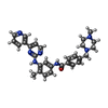
Mass: 493.603 Da / Num. of mol.: 3 / Source method: obtained synthetically / Formula: C29H31N7O / Feature type: SUBJECT OF INVESTIGATION / Comment: medication, inhibitor*YM
#3: Chemical ChemComp-1N1 / N-(2-CHLORO-6-METHYLPHENYL)-2-({6-[4-(2-HYDROXYETHYL)PIPERAZIN-1-YL]-2-METHYLPYRIMIDIN-4-YL}AMINO)-1,3-THIAZOLE-5-CARBOXAMIDE / Dasatinib


Mass: 488.006 Da / Num. of mol.: 3 / Source method: obtained synthetically / Formula: C22H26ClN7O2S / Feature type: SUBJECT OF INVESTIGATION / Comment: medication*YM
#4: Chemical ChemComp-PO4 / PHOSPHATE ION


Mass: 94.971 Da / Num. of mol.: 1 / Source method: obtained synthetically / Formula: PO4
#5: Water ChemComp-HOH / water


Mass: 18.015 Da / Num. of mol.: 206 / Source method: isolated from a natural source / Formula: H2O
Has ligand of interestY

-
Experimental details

-
Experiment

ExperimentMethod: X-RAY DIFFRACTION / Number of used crystals: 1

-
Sample preparation

CrystalDensity Matthews: 2.88 Å3/Da / Density % sol: 57.36 %
Crystal growTemperature: 277.15 K / Method: vapor diffusion, sitting drop / Details: Ammonium dihydrogen phosphate

-
Data collection

DiffractionMean temperature: 100 K / Serial crystal experiment: N
Diffraction sourceSource: SYNCHROTRON / Site: APS / Beamline: 22-BM / Wavelength: 1 Å
DetectorType: RAYONIX MX300-HS / Detector: CCD / Date: Mar 31, 2018
RadiationProtocol: SINGLE WAVELENGTH / Monochromatic (M) / Laue (L): M / Scattering type: x-ray
Radiation wavelengthWavelength: 1 Å / Relative weight: 1
ReflectionResolution: 2.2→46.7 Å / Num. obs: 55951 / % possible obs: 99.9 % / Observed criterion σ(I): -3 / Redundancy: 8.6 % / CC1/2: 0.999 / Rpim(I) all: 0.03 / Rsym value: 0.083 / Net I/σ(I): 15.1
Reflection shellResolution: 2.2→2.26 Å / Redundancy: 8.7 % / Num. unique obs: 4480 / CC1/2: 0.762 / Rsym value: 1.184 / % possible all: 98.6

-
Processing

Software
NameVersionClassification
REFMAC5.8.0257refinement
XDSdata reduction
Aimlessdata scaling
PHASERphasing
RefinementMethod to determine structure: MOLECULAR REPLACEMENT
Starting model: 2GQG
Resolution: 2.2→46.7 Å / Cor.coef. Fo:Fc: 0.948 / Cor.coef. Fo:Fc free: 0.939 / SU B: 7.206 / SU ML: 0.175 / Cross valid method: THROUGHOUT / ESU R: 0.231 / ESU R Free: 0.193
Details: Hydrogens have been added in their riding positions
RfactorNum. reflection% reflection
Rfree0.2511 2704 4.834 %
Rwork0.2174 53232 -
all0.219 --
obs-55936 99.759 %
Solvent computationIon probe radii: 0.8 Å / Shrinkage radii: 0.8 Å / VDW probe radii: 1.2 Å / Solvent model: MASK BULK SOLVENT
Displacement parametersBiso mean: 52.067 Å2
Baniso -1Baniso -2Baniso -3
1-3.585 Å2-0 Å2-0 Å2
2---1.887 Å2-0 Å2
3----1.698 Å2
Refinement stepCycle: LAST / Resolution: 2.2→46.7 Å
ProteinNucleic acidLigandSolventTotal
Num. atoms5886 0 215 206 6307
Refine LS restraints
Refine-IDTypeDev idealDev ideal targetNumber
X-RAY DIFFRACTIONr_bond_refined_d0.0050.0136264
X-RAY DIFFRACTIONr_bond_other_d0.0010.0175577
X-RAY DIFFRACTIONr_angle_refined_deg1.2891.6448506
X-RAY DIFFRACTIONr_angle_other_deg1.2441.5712872
X-RAY DIFFRACTIONr_dihedral_angle_1_deg6.7125736
X-RAY DIFFRACTIONr_dihedral_angle_2_deg34.25522.91299
X-RAY DIFFRACTIONr_dihedral_angle_3_deg14.4115978
X-RAY DIFFRACTIONr_dihedral_angle_4_deg11.5061527
X-RAY DIFFRACTIONr_chiral_restr0.0560.2776
X-RAY DIFFRACTIONr_gen_planes_refined0.0050.026973
X-RAY DIFFRACTIONr_gen_planes_other0.0010.021361
X-RAY DIFFRACTIONr_nbd_refined0.1960.21178
X-RAY DIFFRACTIONr_symmetry_nbd_other0.1760.24971
X-RAY DIFFRACTIONr_nbtor_refined0.170.23066
X-RAY DIFFRACTIONr_symmetry_nbtor_other0.0810.22464
X-RAY DIFFRACTIONr_xyhbond_nbd_refined0.1390.2196
X-RAY DIFFRACTIONr_symmetry_nbd_refined0.1590.210
X-RAY DIFFRACTIONr_nbd_other0.1950.235
X-RAY DIFFRACTIONr_symmetry_xyhbond_nbd_refined0.1450.28
X-RAY DIFFRACTIONr_mcbond_it3.175.6482980
X-RAY DIFFRACTIONr_mcbond_other3.175.6472978
X-RAY DIFFRACTIONr_mcangle_it4.7418.4463702
X-RAY DIFFRACTIONr_mcangle_other4.7418.4473703
X-RAY DIFFRACTIONr_scbond_it3.8455.8343282
X-RAY DIFFRACTIONr_scbond_other3.8435.8363279
X-RAY DIFFRACTIONr_scangle_it5.9098.6274803
X-RAY DIFFRACTIONr_scangle_other5.9088.6314798
X-RAY DIFFRACTIONr_lrange_it7.47462.8317051
X-RAY DIFFRACTIONr_lrange_other7.47362.8117035
LS refinement shell
Resolution (Å)Rfactor RfreeNum. reflection RfreeRfactor RworkNum. reflection RworkRefine-ID% reflection obs (%)
2.2-2.2550.311940.3063811X-RAY DIFFRACTION98.2099
2.255-2.3160.3152160.33794X-RAY DIFFRACTION99.9751
2.316-2.3840.3081740.2623684X-RAY DIFFRACTION99.9223
2.384-2.4570.3011950.2543588X-RAY DIFFRACTION99.9208
2.457-2.5370.3031820.2483463X-RAY DIFFRACTION100
2.537-2.6260.2931750.2493387X-RAY DIFFRACTION100
2.626-2.7260.2641530.2273285X-RAY DIFFRACTION99.9419
2.726-2.8370.2861630.2123125X-RAY DIFFRACTION100
2.837-2.9630.2581580.2143025X-RAY DIFFRACTION100
2.963-3.1070.31460.222867X-RAY DIFFRACTION99.8674
3.107-3.2750.2611410.2072758X-RAY DIFFRACTION99.9311
3.275-3.4740.2391170.2122636X-RAY DIFFRACTION99.8549
3.474-3.7140.2461300.212459X-RAY DIFFRACTION99.8457
3.714-4.0110.2141070.1982264X-RAY DIFFRACTION99.6637
4.011-4.3930.209920.1782160X-RAY DIFFRACTION99.8227
4.393-4.9110.206920.1841923X-RAY DIFFRACTION99.9008
4.911-5.6690.273990.2121709X-RAY DIFFRACTION100
5.669-6.940.289740.241462X-RAY DIFFRACTION99.9349
6.94-9.7990.204570.1921157X-RAY DIFFRACTION100

+
About Yorodumi

-
News

-
Feb 9, 2022. New format data for meta-information of EMDB entries

New format data for meta-information of EMDB entries

  • Version 3 of the EMDB header file is now the official format.
  • The previous official version 1.9 will be removed from the archive.

Related info.:EMDB header

External links:wwPDB to switch to version 3 of the EMDB data model

-
Aug 12, 2020. Covid-19 info

Covid-19 info

URL: https://pdbj.org/emnavi/covid19.php

New page: Covid-19 featured information page in EM Navigator.

Related info.:Covid-19 info / Mar 5, 2020. Novel coronavirus structure data

+
Mar 5, 2020. Novel coronavirus structure data

Novel coronavirus structure data

Related info.:Yorodumi Speices / Aug 12, 2020. Covid-19 info

External links:COVID-19 featured content - PDBj / Molecule of the Month (242):Coronavirus Proteases

+
Jan 31, 2019. EMDB accession codes are about to change! (news from PDBe EMDB page)

EMDB accession codes are about to change! (news from PDBe EMDB page)

  • The allocation of 4 digits for EMDB accession codes will soon come to an end. Whilst these codes will remain in use, new EMDB accession codes will include an additional digit and will expand incrementally as the available range of codes is exhausted. The current 4-digit format prefixed with “EMD-” (i.e. EMD-XXXX) will advance to a 5-digit format (i.e. EMD-XXXXX), and so on. It is currently estimated that the 4-digit codes will be depleted around Spring 2019, at which point the 5-digit format will come into force.
  • The EM Navigator/Yorodumi systems omit the EMD- prefix.

Related info.:Q: What is EMD? / ID/Accession-code notation in Yorodumi/EM Navigator

External links:EMDB Accession Codes are Changing Soon! / Contact to PDBj

+
Jul 12, 2017. Major update of PDB

Major update of PDB

  • wwPDB released updated PDB data conforming to the new PDBx/mmCIF dictionary.
  • This is a major update changing the version number from 4 to 5, and with Remediation, in which all the entries are updated.
  • In this update, many items about electron microscopy experimental information are reorganized (e.g. em_software).
  • Now, EM Navigator and Yorodumi are based on the updated data.

External links:wwPDB Remediation / Enriched Model Files Conforming to OneDep Data Standards Now Available in the PDB FTP Archive

-
Yorodumi

Thousand views of thousand structures

  • Yorodumi is a browser for structure data from EMDB, PDB, SASBDB, etc.
  • This page is also the successor to EM Navigator detail page, and also detail information page/front-end page for Omokage search.
  • The word "yorodu" (or yorozu) is an old Japanese word meaning "ten thousand". "mi" (miru) is to see.

Related info.:EMDB / PDB / SASBDB / Comparison of 3 databanks / Yorodumi Search / Aug 31, 2016. New EM Navigator & Yorodumi / Yorodumi Papers / Jmol/JSmol / Function and homology information / Changes in new EM Navigator and Yorodumi

Read more