long-chain fatty acid import into cell / negative regulation of TORC2 signaling / cellular response to nutrient / TORC1 signaling / phosphatidylinositol-mediated signaling / germ cell development / TOR signaling / positive regulation of translational initiation / mTORC1-mediated signalling / cellular response to dexamethasone stimulus ...long-chain fatty acid import into cell / negative regulation of TORC2 signaling / cellular response to nutrient / TORC1 signaling / phosphatidylinositol-mediated signaling / germ cell development / TOR signaling / positive regulation of translational initiation / mTORC1-mediated signalling / cellular response to dexamethasone stimulus / behavioral fear response / positive regulation of TORC1 signaling / protein serine/threonine/tyrosine kinase activity / negative regulation of insulin receptor signaling pathway / positive regulation of mitotic cell cycle / positive regulation of translation / negative regulation of extrinsic apoptotic signaling pathway / peptidyl-serine phosphorylation / G1/S transition of mitotic cell cycle / response to nutrient levels / modulation of chemical synaptic transmission / cellular response to type II interferon / cellular response to growth factor stimulus / cellular response to insulin stimulus / mitochondrial outer membrane / protein kinase activity / non-specific serine/threonine protein kinase / neuron projection / protein serine kinase activity / protein serine/threonine kinase activity / apoptotic process / negative regulation of apoptotic process / glutamatergic synapse / signal transduction / mitochondrion / nucleoplasm / ATP binding / nucleus / cytoplasm / cytosol Similarity search - Function
Ribosomal protein S6 kinase / Protein kinase, C-terminal / Protein kinase C terminal domain / Extension to Ser/Thr-type protein kinases / AGC-kinase, C-terminal / AGC-kinase C-terminal domain profile. / Transferase(Phosphotransferase) domain 1 / Transferase(Phosphotransferase); domain 1 / Serine/threonine-protein kinase, active site / Serine/Threonine protein kinases active-site signature. ...Ribosomal protein S6 kinase / Protein kinase, C-terminal / Protein kinase C terminal domain / Extension to Ser/Thr-type protein kinases / AGC-kinase, C-terminal / AGC-kinase C-terminal domain profile. / Transferase(Phosphotransferase) domain 1 / Transferase(Phosphotransferase); domain 1 / Serine/threonine-protein kinase, active site / Serine/Threonine protein kinases active-site signature. / Protein kinase domain / Serine/Threonine protein kinases, catalytic domain / Protein kinase, ATP binding site / Protein kinases ATP-binding region signature. / Protein kinase domain profile. / Protein kinase domain / Protein kinase-like domain superfamily / Orthogonal Bundle / Mainly Alpha Similarity search - Domain/homology
Resolution: 2.74→20 Å / Cor.coef. Fo:Fc: 0.946 / Cor.coef. Fo:Fc free: 0.907 / SU B: 38.003 / SU ML: 0.36 / Cross valid method: THROUGHOUT / σ(F): 0 / ESU R Free: 0.43 / Stereochemistry target values: MAXIMUM LIKELIHOOD Details: HYDROGENS HAVE BEEN ADDED IN THE RIDING POSITIONS U VALUES : RESIDUAL ONLY
Rfactor
Num. reflection
% reflection
Selection details
Rfree
0.29
941
5 %
RANDOM
Rwork
0.2115
-
-
-
obs
0.2155
17873
100 %
-
Solvent computation
Ion probe radii: 0.8 Å / Shrinkage radii: 0.8 Å / VDW probe radii: 1.4 Å / Solvent model: MASK
In the structure databanks used in Yorodumi, some data are registered as the other names, "COVID-19 virus" and "2019-nCoV". Here are the details of the virus and the list of structure data.
Jan 31, 2019. EMDB accession codes are about to change! (news from PDBe EMDB page)
EMDB accession codes are about to change! (news from PDBe EMDB page)
The allocation of 4 digits for EMDB accession codes will soon come to an end. Whilst these codes will remain in use, new EMDB accession codes will include an additional digit and will expand incrementally as the available range of codes is exhausted. The current 4-digit format prefixed with “EMD-” (i.e. EMD-XXXX) will advance to a 5-digit format (i.e. EMD-XXXXX), and so on. It is currently estimated that the 4-digit codes will be depleted around Spring 2019, at which point the 5-digit format will come into force.
The EM Navigator/Yorodumi systems omit the EMD- prefix.
Related info.:Q: What is EMD? / ID/Accession-code notation in Yorodumi/EM Navigator
Yorodumi is a browser for structure data from EMDB, PDB, SASBDB, etc.
This page is also the successor to EM Navigator detail page, and also detail information page/front-end page for Omokage search.
The word "yorodu" (or yorozu) is an old Japanese word meaning "ten thousand". "mi" (miru) is to see.
Related info.:EMDB / PDB / SASBDB / Comparison of 3 databanks / Yorodumi Search / Aug 31, 2016. New EM Navigator & Yorodumi / Yorodumi Papers / Jmol/JSmol / Function and homology information / Changes in new EM Navigator and Yorodumi
 Movie
Movie Controller


 Open data
 Basic information
 Components
 Keywords
 Function and homology information
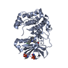
 Homo sapiens (human)
 X-RAY DIFFRACTION /
  Authors
 Citation
 Structure visualization
 Downloads & links
 Other downloads
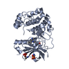
 PDBj
PDBj


 Assembly
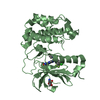

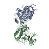
 Spodoptera frugiperda (fall armyworm)
 Mass: 449.857 Da / Num. of mol.: 2 / Source method: obtained synthetically / Formula: C21H19ClF3N5O / Feature type: SUBJECT OF INVESTIGATION
 Mass: 18.015 Da / Num. of mol.: 4 / Source method: isolated from a natural source / Formula: H2O
 Sample preparation
 / Beamline: X06DA / Wavelength: 1 Å
 Processing