 Movie
Movie Controller

[English] 日本語
 Yorodumi
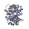
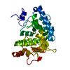
- PDB-7n7i: X-ray crystal structure of Viperin-like enzyme from Trichoderma virens -
+ Open data

- Basic information
| Entry | Database: PDB / ID: 7n7i | ||||||||||||||||||
|---|---|---|---|---|---|---|---|---|---|---|---|---|---|---|---|---|---|---|---|
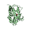
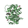
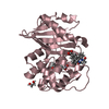
| Title | X-ray crystal structure of Viperin-like enzyme from Trichoderma virens | ||||||||||||||||||
 Components Components | Viperin-like enzyme | ||||||||||||||||||
 Keywords Keywords | ANTIVIRAL PROTEIN / radical SAM protein / metalloprotein / antiviral / ddh-synthase | ||||||||||||||||||
| Function / homology |  Function and homology information Function and homology informationcatalytic activity / 4 iron, 4 sulfur cluster binding / defense response to virus / nucleotide binding / metal ion binding Similarity search - Function | ||||||||||||||||||
| Biological species |  Hypocrea virens (fungus) Hypocrea virens (fungus) | ||||||||||||||||||
| Method |  X-RAY DIFFRACTION / SYNCHROTRON / MOLECULAR REPLACEMENT / Resolution: 3.19 Å X-RAY DIFFRACTION / SYNCHROTRON / MOLECULAR REPLACEMENT / Resolution: 3.19 Å | ||||||||||||||||||
 Authors Authors | Grove, T.L. / Almo, S.C. / Bonanno, J.B. / Lachowicz, J.C. / Gizzi, A.G. | ||||||||||||||||||
| Funding support |  United States, 5items United States, 5items
| ||||||||||||||||||
 Citation Citation | Journal: Biochemistry / Year: 2021 Title: Structural Insight into the Substrate Scope of Viperin and Viperin-like Enzymes from Three Domains of Life. Authors: Lachowicz, J.C. / Gizzi, A.S. / Almo, S.C. / Grove, T.L. | ||||||||||||||||||
| History |
|
- Structure visualization
| Structure viewer | Molecule: MolmilJmol/JSmol |
|---|
- Downloads & links
-Download
| PDBx/mmCIF format | 7n7i.cif.gz | 186.3 KB | Display | PDBx/mmCIF format |
|---|---|---|---|---|
| PDB format | pdb7n7i.ent.gz | 144.9 KB | Display | PDB format |
| PDBx/mmJSON format | 7n7i.json.gz | Tree view | PDBx/mmJSON format | |
| Others |  Other downloads Other downloads |
-Validation report
| Arichive directory | https://data.pdbj.org/pub/pdb/validation_reports/n7/7n7iftp://data.pdbj.org/pub/pdb/validation_reports/n7/7n7i | HTTPS FTP |
|---|
-Related structure data
| Related structure data | 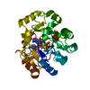 7n7hC 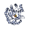 5vslS S: Starting model for refinement C: citing same article ( |
|---|---|
| Similar structure data |


-Links
 PDBj
PDBj
- Assembly
| Deposited unit | 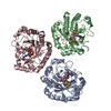
| ||||||||||||||||||||||||||||
|---|---|---|---|---|---|---|---|---|---|---|---|---|---|---|---|---|---|---|---|---|---|---|---|---|---|---|---|---|---|
| 1 | 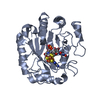
| ||||||||||||||||||||||||||||
| 2 | 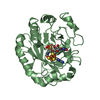
| ||||||||||||||||||||||||||||
| 3 | 
| ||||||||||||||||||||||||||||
| Unit cell |
| ||||||||||||||||||||||||||||
| Noncrystallographic symmetry (NCS) | NCS domain:
NCS domain segments: Component-ID: 1 / Ens-ID: 1 / Beg auth comp-ID: GLN / Beg label comp-ID: GLN / End auth comp-ID: SER / End label comp-ID: SER / Auth seq-ID: 18 - 298 / Label seq-ID: 40 - 320
|
-Components
| #1: Protein | Mass: 37797.598 Da / Num. of mol.: 3 Source method: isolated from a genetically manipulated source Source: (gene. exp.) Hypocrea virens (strain Gv29-8 / FGSC 10586) (fungus)Strain: Gv29-8 / FGSC 10586 / Gene: TRIVIDRAFT_58105 / Production host:  #2: Chemical |   Mass: 398.437 Da / Num. of mol.: 3 / Source method: obtained synthetically / Formula: C15H22N6O5S / Feature type: SUBJECT OF INVESTIGATION Mass: 398.437 Da / Num. of mol.: 3 / Source method: obtained synthetically / Formula: C15H22N6O5S / Feature type: SUBJECT OF INVESTIGATION#3: Chemical |   Mass: 351.640 Da / Num. of mol.: 3 / Source method: obtained synthetically / Formula: Fe4S4 / Feature type: SUBJECT OF INVESTIGATION Mass: 351.640 Da / Num. of mol.: 3 / Source method: obtained synthetically / Formula: Fe4S4 / Feature type: SUBJECT OF INVESTIGATION#4: Chemical |   Mass: 484.141 Da / Num. of mol.: 3 / Source method: obtained synthetically / Formula: C9H15N2O15P3 / Feature type: SUBJECT OF INVESTIGATION / Comment: UTP*YM Mass: 484.141 Da / Num. of mol.: 3 / Source method: obtained synthetically / Formula: C9H15N2O15P3 / Feature type: SUBJECT OF INVESTIGATION / Comment: UTP*YMHas ligand of interest | Y | |
|---|
-Experimental details
-Experiment
| Experiment | Method: X-RAY DIFFRACTION / Number of used crystals: 1 |
|---|
- Sample preparation
| Crystal | Density Matthews: 2.1 Å3/Da / Density % sol: 41.54 % |
|---|---|
| Crystal grow | Temperature: 298 K / Method: vapor diffusion, sitting drop / pH: 8.5 Details: 0.1 M Tris-HCl, pH 8.5, 0.2 M sodium chloride, 25% (w/v) PEG 3350 |
-Data collection
| Diffraction | Mean temperature: 100 K / Serial crystal experiment: N | ||||||||||||||||||||||||
|---|---|---|---|---|---|---|---|---|---|---|---|---|---|---|---|---|---|---|---|---|---|---|---|---|---|
| Diffraction source | Source: SYNCHROTRON / Site: NSLS-II / Beamline: 17-ID-2 / Wavelength: 0.9893 Å | ||||||||||||||||||||||||
| Detector | Type: DECTRIS EIGER X 16M / Detector: PIXEL / Date: Dec 7, 2017 | ||||||||||||||||||||||||
| Radiation | Protocol: SINGLE WAVELENGTH / Monochromatic (M) / Laue (L): M / Scattering type: x-ray | ||||||||||||||||||||||||
| Radiation wavelength | Wavelength: 0.9893 Å / Relative weight: 1 | ||||||||||||||||||||||||
| Reflection | Resolution: 3.19→28.14 Å / Num. obs: 15361 / % possible obs: 98.8 % / Redundancy: 10.3 % / CC1/2: 0.982 / Rmerge(I) obs: 0.2 / Rpim(I) all: 0.129 / Rrim(I) all: 0.409 / Net I/σ(I): 8.46 | ||||||||||||||||||||||||
| Reflection shell | Diffraction-ID: 1
|
- Processing
| Software |
| ||||||||||||||||||||||||||||||||||||||||||||||||||||||||||||||||||||||||||||||||||||
|---|---|---|---|---|---|---|---|---|---|---|---|---|---|---|---|---|---|---|---|---|---|---|---|---|---|---|---|---|---|---|---|---|---|---|---|---|---|---|---|---|---|---|---|---|---|---|---|---|---|---|---|---|---|---|---|---|---|---|---|---|---|---|---|---|---|---|---|---|---|---|---|---|---|---|---|---|---|---|---|---|---|---|---|---|---|
| Refinement | Method to determine structure: MOLECULAR REPLACEMENT Starting model: 5VSL Resolution: 3.19→28.14 Å / SU ML: 0.47 / Cross valid method: THROUGHOUT / σ(F): 0.11 / Phase error: 26.25 / Stereochemistry target values: ML
| ||||||||||||||||||||||||||||||||||||||||||||||||||||||||||||||||||||||||||||||||||||
| Solvent computation | Shrinkage radii: 0.9 Å / VDW probe radii: 1.11 Å / Solvent model: FLAT BULK SOLVENT MODEL | ||||||||||||||||||||||||||||||||||||||||||||||||||||||||||||||||||||||||||||||||||||
| Displacement parameters | Biso max: 118.75 Å2 / Biso mean: 42.9874 Å2 / Biso min: 13.3 Å2 | ||||||||||||||||||||||||||||||||||||||||||||||||||||||||||||||||||||||||||||||||||||
| Refinement step | Cycle: final / Resolution: 3.19→28.14 Å
| ||||||||||||||||||||||||||||||||||||||||||||||||||||||||||||||||||||||||||||||||||||
| Refine LS restraints NCS |
| ||||||||||||||||||||||||||||||||||||||||||||||||||||||||||||||||||||||||||||||||||||
| LS refinement shell | Refine-ID: X-RAY DIFFRACTION / Rfactor Rfree error: 0 / Total num. of bins used: 11
|
