| Deposited unit | A: Isoform 2 of Chromodomain Y-like protein
G: inhibitor UNC6261
B: Isoform 2 of Chromodomain Y-like protein
H: inhibitor UNC6261
C: Isoform 2 of Chromodomain Y-like protein
I: inhibitor UNC6261
D: Isoform 2 of Chromodomain Y-like protein
J: inhibitor UNC6261
E: Isoform 2 of Chromodomain Y-like protein
K: inhibitor UNC6261
F: Isoform 2 of Chromodomain Y-like protein
L: inhibitor UNC6261
hetero molecules
| Theoretical mass | Number of molelcules |
|---|
| Total (without water) | 48,321 | 16 |
|---|
| Polymers | 48,275 | 12 |
|---|
| Non-polymers | 46 | 4 |
|---|
| Water | 1,243 | 69 |
|---|
|
|---|
| 1 | A: Isoform 2 of Chromodomain Y-like protein
G: inhibitor UNC6261
hetero molecules
| Theoretical mass | Number of molelcules |
|---|
| Total (without water) | 8,069 | 3 |
|---|
| Polymers | 8,046 | 2 |
|---|
| Non-polymers | 23 | 1 |
|---|
| Water | 36 | 2 |
|---|
| Type | Name | Symmetry operation | Number |
|---|
| identity operation | 1_555 | x,y,z | 1 |
| Buried area | 1410 Å2 |
|---|
| ΔGint | -2 kcal/mol |
|---|
| Surface area | 4360 Å2 |
|---|
| Method | PISA |
|---|
|
|---|
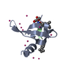
| 2 | B: Isoform 2 of Chromodomain Y-like protein
H: inhibitor UNC6261
| Theoretical mass | Number of molelcules |
|---|
| Total (without water) | 8,046 | 2 |
|---|
| Polymers | 8,046 | 2 |
|---|
| Non-polymers | 0 | 0 |
|---|
| Water | 36 | 2 |
|---|
| Type | Name | Symmetry operation | Number |
|---|
| identity operation | 1_555 | x,y,z | 1 |
| Buried area | 1430 Å2 |
|---|
| ΔGint | -1 kcal/mol |
|---|
| Surface area | 4500 Å2 |
|---|
| Method | PISA |
|---|
|
|---|
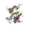
| 3 | C: Isoform 2 of Chromodomain Y-like protein
I: inhibitor UNC6261
| Theoretical mass | Number of molelcules |
|---|
| Total (without water) | 8,046 | 2 |
|---|
| Polymers | 8,046 | 2 |
|---|
| Non-polymers | 0 | 0 |
|---|
| Water | 18 | 1 |
|---|
| Type | Name | Symmetry operation | Number |
|---|
| identity operation | 1_555 | x,y,z | 1 |
| Buried area | 1500 Å2 |
|---|
| ΔGint | -10 kcal/mol |
|---|
| Surface area | 4490 Å2 |
|---|
| Method | PISA |
|---|
|
|---|
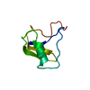
| 4 | D: Isoform 2 of Chromodomain Y-like protein
J: inhibitor UNC6261
hetero molecules
| Theoretical mass | Number of molelcules |
|---|
| Total (without water) | 8,069 | 4 |
|---|
| Polymers | 8,046 | 2 |
|---|
| Non-polymers | 23 | 2 |
|---|
| Water | 36 | 2 |
|---|
| Type | Name | Symmetry operation | Number |
|---|
| identity operation | 1_555 | x,y,z | 1 |
| Buried area | 1520 Å2 |
|---|
| ΔGint | -9 kcal/mol |
|---|
| Surface area | 4700 Å2 |
|---|
| Method | PISA |
|---|
|
|---|
| 5 | E: Isoform 2 of Chromodomain Y-like protein
K: inhibitor UNC6261
| Theoretical mass | Number of molelcules |
|---|
| Total (without water) | 8,046 | 2 |
|---|
| Polymers | 8,046 | 2 |
|---|
| Non-polymers | 0 | 0 |
|---|
| Water | 36 | 2 |
|---|
| Type | Name | Symmetry operation | Number |
|---|
| identity operation | 1_555 | x,y,z | 1 |
| Buried area | 1340 Å2 |
|---|
| ΔGint | -1 kcal/mol |
|---|
| Surface area | 4290 Å2 |
|---|
| Method | PISA |
|---|
|
|---|
| 6 | F: Isoform 2 of Chromodomain Y-like protein
L: inhibitor UNC6261
| Theoretical mass | Number of molelcules |
|---|
| Total (without water) | 8,046 | 3 |
|---|
| Polymers | 8,046 | 2 |
|---|
| Non-polymers | 0 | 1 |
|---|
| Water | 36 | 2 |
|---|
| Type | Name | Symmetry operation | Number |
|---|
| identity operation | 1_555 | x,y,z | 1 |
| Buried area | 1380 Å2 |
|---|
| ΔGint | -1 kcal/mol |
|---|
| Surface area | 4610 Å2 |
|---|
| Method | PISA |
|---|
|
|---|
| Unit cell | | Length a, b, c (Å) | 62.971, 76.386, 80.628 |
|---|
| Angle α, β, γ (deg.) | 90.00, 90.00, 90.00 |
|---|
| Int Tables number | 19 |
|---|
| Space group name H-M | P212121 |
|---|
|
|---|
 Movie
Movie Controller

 Yorodumi
 Open data
 Basic information
 Components
 Keywords
 Function and homology information
 Homo sapiens (human)
 X-RAY DIFFRACTION /
  Authors
 Citation
 Structure visualization
 Downloads & links
 Other downloads
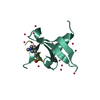


 PDBj
PDBj Assembly


 Mass: 22.990 Da / Num. of mol.: 2 / Source method: obtained synthetically / Formula: Na
 Num. of mol.: 2 / Source method: obtained synthetically
 Mass: 18.015 Da / Num. of mol.: 69 / Source method: isolated from a natural source / Formula: H2O
 Sample preparation
 / Beamline: 24-ID-E / Wavelength: 0.97934 Å
 Processing