 Movie
Movie Controller


+ Open data

- Basic information
| Entry | Database: PDB / ID: 7my1 | ||||||
|---|---|---|---|---|---|---|---|
| Title | Sy-CrtE structure with IPP, N-term His-tag | ||||||
 Components Components | Geranylgeranyl pyrophosphate synthase | ||||||
 Keywords Keywords | TRANSFERASE / Prenyl transferase / Isopentenyl pyrophosphate / Dimethylallyl pyrophosphate / cyanobacteria / terpenoids | ||||||
| Function / homology |  Function and homology information Function and homology informationdimethylallyltranstransferase / prenyltransferase activity / isoprenoid biosynthetic process / terpenoid biosynthetic process / dimethylallyltranstransferase activity / metal ion binding / cytoplasm Similarity search - Function | ||||||
| Biological species |  | ||||||
| Method |  X-RAY DIFFRACTION / SYNCHROTRON / MOLECULAR REPLACEMENT / Resolution: 1.84 Å X-RAY DIFFRACTION / SYNCHROTRON / MOLECULAR REPLACEMENT / Resolution: 1.84 Å | ||||||
 Authors Authors | Peat, T.S. / Newman, J. | ||||||
 Citation Citation | Journal: Febs J. / Year: 2022 Title: Molecular characterization of cyanobacterial short-chain prenyltransferases and discovery of a novel GGPP phosphatase. Authors: Satta, A. / Esquirol, L. / Ebert, B.E. / Newman, J. / Peat, T.S. / Plan, M. / Schenk, G. / Vickers, C.E. | ||||||
| History |
|
- Structure visualization
| Structure viewer | Molecule: MolmilJmol/JSmol |
|---|
- Downloads & links
-Download
| PDBx/mmCIF format | 7my1.cif.gz | 77.1 KB | Display | PDBx/mmCIF format |
|---|---|---|---|---|
| PDB format | pdb7my1.ent.gz | Display | PDB format | |
| PDBx/mmJSON format | 7my1.json.gz | Tree view | PDBx/mmJSON format | |
| Others |  Other downloads Other downloads |
-Validation report
| Arichive directory | https://data.pdbj.org/pub/pdb/validation_reports/my/7my1ftp://data.pdbj.org/pub/pdb/validation_reports/my/7my1 | HTTPS FTP |
|---|
-Related structure data
| Related structure data | 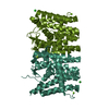 7mxzSC 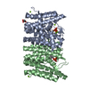 7my0C 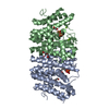 7my6C 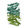 7my7C S: Starting model for refinement C: citing same article ( |
|---|---|
| Similar structure data |
-Links
 PDBj
PDBj
- Assembly
| Deposited unit | 
| ||||||||||||
|---|---|---|---|---|---|---|---|---|---|---|---|---|---|
| 1 | 
| ||||||||||||
| Unit cell |
| ||||||||||||
| Components on special symmetry positions |
|
-Components
| #1: Protein | Mass: 34707.492 Da / Num. of mol.: 1 Source method: isolated from a genetically manipulated source Source: (gene. exp.) Strain: PCC 6803 / Kazusa / Gene: crtE / Production host: | ||||||
|---|---|---|---|---|---|---|---|
| #2: Chemical | ChemComp-IPE / 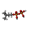  Mass: 246.092 Da / Num. of mol.: 1 / Source method: obtained synthetically / Formula: C5H12O7P2 / Feature type: SUBJECT OF INVESTIGATION Mass: 246.092 Da / Num. of mol.: 1 / Source method: obtained synthetically / Formula: C5H12O7P2 / Feature type: SUBJECT OF INVESTIGATION | ||||||
| #3: Chemical | ChemComp-MG /   Mass: 24.305 Da / Num. of mol.: 4 / Source method: obtained synthetically / Formula: Mg Mass: 24.305 Da / Num. of mol.: 4 / Source method: obtained synthetically / Formula: Mg#4: Chemical | ChemComp-CL / |   Mass: 35.453 Da / Num. of mol.: 1 / Source method: obtained synthetically / Formula: Cl Mass: 35.453 Da / Num. of mol.: 1 / Source method: obtained synthetically / Formula: Cl#5: Water | ChemComp-HOH / |  Mass: 18.015 Da / Num. of mol.: 111 / Source method: isolated from a natural source / Formula: H2O Mass: 18.015 Da / Num. of mol.: 111 / Source method: isolated from a natural source / Formula: H2OHas ligand of interest | Y | |
-Experimental details
-Experiment
| Experiment | Method: X-RAY DIFFRACTION / Number of used crystals: 1 |
|---|
- Sample preparation
| Crystal | Density Matthews: 2.08 Å3/Da / Density % sol: 40.94 % |
|---|---|
| Crystal grow | Temperature: 293 K / Method: vapor diffusion, sitting drop Details: Protein was at 10 mg/mL and the drops were 200 nL plus 200 nL with the reservoir being 22.5% PEG 3350, 0.3M MgCl2, 0.1M Na-HEPES pH 6.55 |
-Data collection
| Diffraction | Mean temperature: 100 K / Serial crystal experiment: N |
|---|---|
| Diffraction source | Source: SYNCHROTRON / Site: Australian Synchrotron  / Beamline: MX2 / Wavelength: 0.9537 Å / Beamline: MX2 / Wavelength: 0.9537 Å |
| Detector | Type: DECTRIS EIGER X 16M / Detector: PIXEL / Date: Nov 14, 2020 |
| Radiation | Protocol: SINGLE WAVELENGTH / Monochromatic (M) / Laue (L): M / Scattering type: x-ray |
| Radiation wavelength | Wavelength: 0.9537 Å / Relative weight: 1 |
| Reflection | Resolution: 1.84→45.16 Å / Num. obs: 24629 / % possible obs: 99 % / Redundancy: 6.7 % / CC1/2: 0.998 / Rmerge(I) obs: 0.1 / Rpim(I) all: 0.042 / Net I/σ(I): 9.4 |
| Reflection shell | Resolution: 1.84→1.88 Å / Rmerge(I) obs: 1.367 / Mean I/σ(I) obs: 1.3 / Num. unique obs: 1295 / CC1/2: 0.574 / Rpim(I) all: 0.587 / % possible all: 85.7 |
- Processing
| Software |
| ||||||||||||||||||||||||||||||||||||||||||||||||||||||||||||||||||||||||||||||||||||||||||||||||||||||||||||||||||||||||||||||||||||||||||||||||||||||||||||||||
|---|---|---|---|---|---|---|---|---|---|---|---|---|---|---|---|---|---|---|---|---|---|---|---|---|---|---|---|---|---|---|---|---|---|---|---|---|---|---|---|---|---|---|---|---|---|---|---|---|---|---|---|---|---|---|---|---|---|---|---|---|---|---|---|---|---|---|---|---|---|---|---|---|---|---|---|---|---|---|---|---|---|---|---|---|---|---|---|---|---|---|---|---|---|---|---|---|---|---|---|---|---|---|---|---|---|---|---|---|---|---|---|---|---|---|---|---|---|---|---|---|---|---|---|---|---|---|---|---|---|---|---|---|---|---|---|---|---|---|---|---|---|---|---|---|---|---|---|---|---|---|---|---|---|---|---|---|---|---|---|---|---|
| Refinement | Method to determine structure: MOLECULAR REPLACEMENT Starting model: 7mxz Resolution: 1.84→45.16 Å / Cor.coef. Fo:Fc: 0.969 / Cor.coef. Fo:Fc free: 0.952 / SU B: 4.61 / SU ML: 0.125 / Cross valid method: FREE R-VALUE / ESU R: 0.14 / ESU R Free: 0.135 Details: Hydrogens have been added in their riding positions
| ||||||||||||||||||||||||||||||||||||||||||||||||||||||||||||||||||||||||||||||||||||||||||||||||||||||||||||||||||||||||||||||||||||||||||||||||||||||||||||||||
| Solvent computation | Ion probe radii: 0.8 Å / Shrinkage radii: 0.8 Å / VDW probe radii: 1.2 Å / Solvent model: MASK BULK SOLVENT | ||||||||||||||||||||||||||||||||||||||||||||||||||||||||||||||||||||||||||||||||||||||||||||||||||||||||||||||||||||||||||||||||||||||||||||||||||||||||||||||||
| Displacement parameters | Biso mean: 30.81 Å2
| ||||||||||||||||||||||||||||||||||||||||||||||||||||||||||||||||||||||||||||||||||||||||||||||||||||||||||||||||||||||||||||||||||||||||||||||||||||||||||||||||
| Refinement step | Cycle: LAST / Resolution: 1.84→45.16 Å
| ||||||||||||||||||||||||||||||||||||||||||||||||||||||||||||||||||||||||||||||||||||||||||||||||||||||||||||||||||||||||||||||||||||||||||||||||||||||||||||||||
| Refine LS restraints |
| ||||||||||||||||||||||||||||||||||||||||||||||||||||||||||||||||||||||||||||||||||||||||||||||||||||||||||||||||||||||||||||||||||||||||||||||||||||||||||||||||
| LS refinement shell |
|
