 Movie
Movie Controller

[English] 日本語
 Yorodumi
- PDB-7mxk: Crystal structure of the S/T protein kinase PknG from Corynebacte... -
+ Open data

- Basic information
| Entry | Database: PDB / ID: 7mxk | ||||||
|---|---|---|---|---|---|---|---|
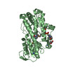
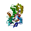
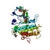
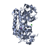
| Title | Crystal structure of the S/T protein kinase PknG from Corynebacterium glutamicum (residues 130-433) in complex with AMP-PNP, isoform 2 | ||||||
 Components Components | Serine/threonine protein kinases | ||||||
 Keywords Keywords | TRANSFERASE / metabolic control / glutamate metabolism / bacterial protein kinase / tetratricopeptide repeats | ||||||
| Function / homology |  Function and homology information Function and homology informationnon-specific serine/threonine protein kinase / protein serine/threonine kinase activity / ATP binding / metal ion binding Similarity search - Function | ||||||
| Biological species |  Corynebacterium glutamicum (bacteria) Corynebacterium glutamicum (bacteria) | ||||||
| Method |  X-RAY DIFFRACTION / SYNCHROTRON / MOLECULAR REPLACEMENT / Resolution: 1.99 Å X-RAY DIFFRACTION / SYNCHROTRON / MOLECULAR REPLACEMENT / Resolution: 1.99 Å | ||||||
 Authors Authors | Lisa, M.N. / Alzari, P.M. | ||||||
| Funding support |  France, 1items France, 1items
| ||||||
 Citation Citation | Journal: Mbio / Year: 2021 Title: A Tetratricopeptide Repeat Scaffold Couples Signal Detection to OdhI Phosphorylation in Metabolic Control by the Protein Kinase PknG. Authors: Lisa, M.N. / Sogues, A. / Barilone, N. / Baumgart, M. / Gil, M. / Grana, M. / Duran, R. / Biondi, R.M. / Bellinzoni, M. / Bott, M. / Alzari, P.M. | ||||||
| History |
|
- Structure visualization
| Structure viewer | Molecule: MolmilJmol/JSmol |
|---|
- Downloads & links
-Download
| PDBx/mmCIF format | 7mxk.cif.gz | 165.6 KB | Display | PDBx/mmCIF format |
|---|---|---|---|---|
| PDB format | pdb7mxk.ent.gz | 107 KB | Display | PDB format |
| PDBx/mmJSON format | 7mxk.json.gz | Tree view | PDBx/mmJSON format | |
| Others |  Other downloads Other downloads |
-Validation report
| Arichive directory | https://data.pdbj.org/pub/pdb/validation_reports/mx/7mxkftp://data.pdbj.org/pub/pdb/validation_reports/mx/7mxk | HTTPS FTP |
|---|
-Related structure data
| Related structure data |  7mxbSC  7mxjC S: Starting model for refinement C: citing same article ( |
|---|---|
| Similar structure data |


-Links
 PDBj
PDBj- Assembly
| Deposited unit | 
| ||||||||||||
|---|---|---|---|---|---|---|---|---|---|---|---|---|---|
| 1 |
| ||||||||||||
| Unit cell |
|
-Components
| #1: Protein | Mass: 33272.176 Da / Num. of mol.: 1 Source method: isolated from a genetically manipulated source Source: (gene. exp.) Corynebacterium glutamicum (strain ATCC 13032 / DSM 20300 / BCRC 11384 / JCM 1318 / LMG 3730 / NCIMB 10025) (bacteria)Strain: ATCC 13032 / DSM 20300 / BCRC 11384 / JCM 1318 / LMG 3730 / NCIMB 10025 Gene: Cgl2751 / Production host: |
|---|---|
| #2: Chemical | ChemComp-MG /   Mass: 24.305 Da / Num. of mol.: 1 / Source method: obtained synthetically / Formula: Mg Mass: 24.305 Da / Num. of mol.: 1 / Source method: obtained synthetically / Formula: Mg |
| #3: Chemical | ChemComp-MAP /   Mass: 529.493 Da / Num. of mol.: 1 / Source method: obtained synthetically / Formula: C10H16MgN6O12P3 / Feature type: SUBJECT OF INVESTIGATION Mass: 529.493 Da / Num. of mol.: 1 / Source method: obtained synthetically / Formula: C10H16MgN6O12P3 / Feature type: SUBJECT OF INVESTIGATION |
| #4: Water | ChemComp-HOH /  Mass: 18.015 Da / Num. of mol.: 157 / Source method: isolated from a natural source / Formula: H2O Mass: 18.015 Da / Num. of mol.: 157 / Source method: isolated from a natural source / Formula: H2O |
| Has ligand of interest | Y |
-Experimental details
-Experiment
| Experiment | Method: X-RAY DIFFRACTION / Number of used crystals: 1 |
|---|
- Sample preparation
| Crystal | Density Matthews: 2.27 Å3/Da / Density % sol: 45.86 % |
|---|---|
| Crystal grow | Temperature: 291 K / Method: vapor diffusion Details: 100 mM Tris-HCl, 27-30% w/v PEG 4 k, 200 mM MgCl2, pH 8.8 |
-Data collection
| Diffraction | Mean temperature: 100 K / Serial crystal experiment: N |
|---|---|
| Diffraction source | Source: SYNCHROTRON / Site: ESRF / Beamline: ID29 / Wavelength: 0.976251 Å |
| Detector | Type: DECTRIS PILATUS 6M / Detector: PIXEL / Date: Feb 22, 2013 |
| Radiation | Protocol: SINGLE WAVELENGTH / Monochromatic (M) / Laue (L): M / Scattering type: x-ray |
| Radiation wavelength | Wavelength: 0.976251 Å / Relative weight: 1 |
| Reflection | Resolution: 1.99→48.83 Å / Num. obs: 21235 / % possible obs: 98.4 % / Redundancy: 6 % / Biso Wilson estimate: 29.95 Å2 / CC1/2: 0.999 / Rmerge(I) obs: 0.067 / Rpim(I) all: 0.029 / Rrim(I) all: 0.073 / Net I/σ(I): 17.1 |
| Reflection shell | Resolution: 1.99→2.04 Å / Redundancy: 5.2 % / Rmerge(I) obs: 0.654 / Num. unique obs: 1207 / CC1/2: 0.751 / Rpim(I) all: 0.306 / Rrim(I) all: 0.725 / % possible all: 84.9 |
- Processing
| Software |
| |||||||||||||||||||||||||||||||||||||||||||||||||||||||||||||||
|---|---|---|---|---|---|---|---|---|---|---|---|---|---|---|---|---|---|---|---|---|---|---|---|---|---|---|---|---|---|---|---|---|---|---|---|---|---|---|---|---|---|---|---|---|---|---|---|---|---|---|---|---|---|---|---|---|---|---|---|---|---|---|---|---|
| Refinement | Method to determine structure: MOLECULAR REPLACEMENT Starting model: 7MXB Resolution: 1.99→43.77 Å / SU ML: 0.222 / Cross valid method: FREE R-VALUE / σ(F): 1.35 / Phase error: 23.3222 Stereochemistry target values: GeoStd + Monomer Library + CDL v1.2
| |||||||||||||||||||||||||||||||||||||||||||||||||||||||||||||||
| Solvent computation | Shrinkage radii: 0.9 Å / VDW probe radii: 1.11 Å / Solvent model: FLAT BULK SOLVENT MODEL | |||||||||||||||||||||||||||||||||||||||||||||||||||||||||||||||
| Displacement parameters | Biso mean: 47.13 Å2 | |||||||||||||||||||||||||||||||||||||||||||||||||||||||||||||||
| Refinement step | Cycle: LAST / Resolution: 1.99→43.77 Å
| |||||||||||||||||||||||||||||||||||||||||||||||||||||||||||||||
| Refine LS restraints |
| |||||||||||||||||||||||||||||||||||||||||||||||||||||||||||||||
| LS refinement shell |
| |||||||||||||||||||||||||||||||||||||||||||||||||||||||||||||||
| Refinement TLS params. | Method: refined / Origin x: -0.113720250801 Å / Origin y: 17.565592884 Å / Origin z: -18.6506305899 Å
| |||||||||||||||||||||||||||||||||||||||||||||||||||||||||||||||
| Refinement TLS group | Selection details: all |
