 Movie
Movie Controller

[English] 日本語
 Yorodumi
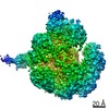
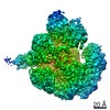
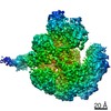
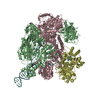
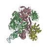
- PDB-7mr3: Cryo-EM structure of RecBCD-DNA complex with docked RecBNuc and s... -
+ Open data

- Basic information
| Entry | Database: PDB / ID: 7mr3 | ||||||||||||||||||||||||||||||||||||
|---|---|---|---|---|---|---|---|---|---|---|---|---|---|---|---|---|---|---|---|---|---|---|---|---|---|---|---|---|---|---|---|---|---|---|---|---|---|
| Title | Cryo-EM structure of RecBCD-DNA complex with docked RecBNuc and stabilized RecD | ||||||||||||||||||||||||||||||||||||
 Components Components |
| ||||||||||||||||||||||||||||||||||||
 Keywords Keywords | HYDROLASE/DNA / SF1 helicase / complex / DNA repair / motor protein / HYDROLASE-DNA complex | ||||||||||||||||||||||||||||||||||||
| Function / homology |  Function and homology information Function and homology informationexodeoxyribonuclease V / exodeoxyribonuclease V activity / exodeoxyribonuclease V complex / clearance of foreign intracellular DNA / DNA translocase activity / DNA 5'-3' helicase / single-stranded DNA helicase activity / recombinational repair / 3'-5' DNA helicase activity / DNA 3'-5' helicase ...exodeoxyribonuclease V / exodeoxyribonuclease V activity / exodeoxyribonuclease V complex / clearance of foreign intracellular DNA / DNA translocase activity / DNA 5'-3' helicase / single-stranded DNA helicase activity / recombinational repair / 3'-5' DNA helicase activity / DNA 3'-5' helicase / ATP-dependent activity, acting on DNA / DNA helicase activity / helicase activity / DNA endonuclease activity / response to radiation / double-strand break repair via homologous recombination / DNA recombination / 5'-3' DNA helicase activity / DNA damage response / magnesium ion binding / ATP hydrolysis activity / DNA binding / ATP binding / cytosol Similarity search - Function | ||||||||||||||||||||||||||||||||||||
| Biological species |  Synthetic construct (others) | ||||||||||||||||||||||||||||||||||||
| Method | ELECTRON MICROSCOPY / single particle reconstruction / cryo EM / Resolution: 3.6 Å | ||||||||||||||||||||||||||||||||||||
 Authors Authors | Hao, L. / Zhang, R. / Lohman, T.M. | ||||||||||||||||||||||||||||||||||||
| Funding support |  United States, 2items United States, 2items
| ||||||||||||||||||||||||||||||||||||
 Citation Citation | Journal: J Mol Biol / Year: 2021 Title: Heterogeneity in E. coli RecBCD Helicase-DNA Binding and Base Pair Melting. Authors: Linxuan Hao / Rui Zhang / Timothy M Lohman / Abstract: E. coli RecBCD, a helicase/nuclease involved in double stranded (ds) DNA break repair, binds to a dsDNA end and melts out several DNA base pairs (bp) using only its binding free energy. We examined ...E. coli RecBCD, a helicase/nuclease involved in double stranded (ds) DNA break repair, binds to a dsDNA end and melts out several DNA base pairs (bp) using only its binding free energy. We examined RecBCD-DNA initiation complexes using thermodynamic and structural approaches. Measurements of enthalpy changes for RecBCD binding to DNA ends possessing pre-melted ssDNA tails of increasing length suggest that RecBCD interacts with ssDNA as long as 17-18 nucleotides and can melt at least 10-11 bp upon binding a blunt DNA end. Cryo-EM structures of RecBCD alone and in complex with a blunt-ended dsDNA show significant conformational heterogeneities associated with the RecB nuclease domain (RecB) and the RecD subunit. In the absence of DNA, 56% of RecBCD molecules show no density for the RecB nuclease domain, RecB, and all RecBCD molecules show only partial density for RecD. DNA binding reduces these conformational heterogeneities, with 63% of the molecules showing density for both RecD and RecB. This suggests that the RecB domain is dynamic and influenced by DNA binding. The major RecBCD-DNA structural class in which RecB is docked onto RecC shows melting of at least 11 bp from a blunt DNA end, much larger than previously observed. A second structural class in which RecB is not docked shows only four bp melted suggesting that RecBCD complexes transition between states with different extents of DNA melting and that the extent of melting regulates initiation of helicase activity. | ||||||||||||||||||||||||||||||||||||
| History |
|
- Structure visualization
| Movie |
Movie viewer |
|---|---|
| Structure viewer | Molecule: MolmilJmol/JSmol |
- Downloads & links
-Download
| PDBx/mmCIF format | 7mr3.cif.gz | 532.3 KB | Display | PDBx/mmCIF format |
|---|---|---|---|---|
| PDB format | pdb7mr3.ent.gz | 423.9 KB | Display | PDB format |
| PDBx/mmJSON format | 7mr3.json.gz | Tree view | PDBx/mmJSON format | |
| Others |  Other downloads Other downloads |
-Validation report
| Arichive directory | https://data.pdbj.org/pub/pdb/validation_reports/mr/7mr3ftp://data.pdbj.org/pub/pdb/validation_reports/mr/7mr3 | HTTPS FTP |
|---|
-Related structure data
| Related structure data | 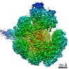 23955MC 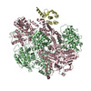 7mr0C 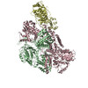 7mr1C 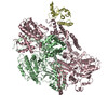 7mr2C 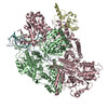 7mr4C M: map data used to model this data C: citing same article ( |
|---|---|
| Similar structure data |


-Links
 PDBj
PDBj


- Assembly
| Deposited unit | 
|
|---|---|
| 1 |
|
-Components
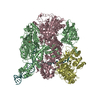
| #1: Protein | Mass: 134110.641 Da / Num. of mol.: 1 Source method: isolated from a genetically manipulated source Source: (gene. exp.) Strain: K12 / Gene: recB, ior, rorA, b2820, JW2788 / Production host: |
|---|---|
| #2: Protein | Mass: 128974.102 Da / Num. of mol.: 1 Source method: isolated from a genetically manipulated source Source: (gene. exp.) Strain: K12 / Gene: recC, b2822, JW2790 / Production host: |
| #3: Protein | Mass: 66990.367 Da / Num. of mol.: 1 Source method: isolated from a genetically manipulated source Source: (gene. exp.) Strain: K12 / Gene: recD, hopE, b2819, JW2787 / Production host: |
| #4: DNA chain | Mass: 18482.836 Da / Num. of mol.: 1 / Source method: obtained synthetically / Source: (synth.) Synthetic construct (others) |
| #5: DNA chain | Mass: 18500.861 Da / Num. of mol.: 1 / Source method: obtained synthetically / Source: (synth.) Synthetic construct (others) |
| Has protein modification | N |
-Experimental details
-Experiment
| Experiment | Method: ELECTRON MICROSCOPY |
|---|---|
| EM experiment | Aggregation state: PARTICLE / 3D reconstruction method: single particle reconstruction |
- Sample preparation
| Component | Name: RecBCD heterotrimer in complex with 60bp blunt-ended dsDNA Type: COMPLEX / Entity ID: #1-#3 / Source: MULTIPLE SOURCES | ||||||||||||||||||||
|---|---|---|---|---|---|---|---|---|---|---|---|---|---|---|---|---|---|---|---|---|---|
| Source (natural) | Organism: | ||||||||||||||||||||
| Source (recombinant) | Organism: | ||||||||||||||||||||
| Buffer solution | pH: 7.4 | ||||||||||||||||||||
| Buffer component |
| ||||||||||||||||||||
| Specimen | Embedding applied: NO / Shadowing applied: NO / Staining applied: NO / Vitrification applied: YES | ||||||||||||||||||||
| Specimen support | Grid material: COPPER / Grid mesh size: 300 divisions/in. / Grid type: Quantifoil R2/2 | ||||||||||||||||||||
| Vitrification | Instrument: FEI VITROBOT MARK IV / Cryogen name: ETHANE / Humidity: 100 % |
- Electron microscopy imaging
| Experimental equipment |  Model: Titan Krios / Image courtesy: FEI Company |
|---|---|
| Microscopy | Model: FEI TITAN KRIOS |
| Electron gun | Electron source:  FIELD EMISSION GUN / Accelerating voltage: 300 kV / Illumination mode: FLOOD BEAM FIELD EMISSION GUN / Accelerating voltage: 300 kV / Illumination mode: FLOOD BEAM |
| Electron lens | Mode: BRIGHT FIELD / Cs: 0.01 mm |
| Image recording | Electron dose: 49.5 e/Å2 / Detector mode: COUNTING / Film or detector model: GATAN K2 QUANTUM (4k x 4k) |
| Image scans | Width: 3838 / Height: 3710 |
- Processing
| Software | Name: PHENIX / Version: 1.19.1_4122: / Classification: refinement | ||||||||||||||||||||||||
|---|---|---|---|---|---|---|---|---|---|---|---|---|---|---|---|---|---|---|---|---|---|---|---|---|---|
| EM software |
| ||||||||||||||||||||||||
| CTF correction | Type: PHASE FLIPPING AND AMPLITUDE CORRECTION | ||||||||||||||||||||||||
| 3D reconstruction | Resolution: 3.6 Å / Resolution method: FSC 0.143 CUT-OFF / Num. of particles: 45202 / Symmetry type: POINT | ||||||||||||||||||||||||
| Refine LS restraints |
|
