 Movie
Movie Controller


+ Open data

- Basic information
| Entry | Database: PDB / ID: 7mm8 | |||||||||
|---|---|---|---|---|---|---|---|---|---|---|
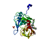
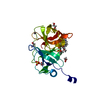
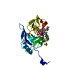
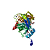
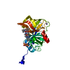
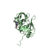
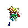
| Title | Crystal structure of HCV NS3/4A protease in complex with NR02-08 | |||||||||
 Components Components | NS3/4a protease | |||||||||
 Keywords Keywords | HYDROLASE/INHIBITOR / NS3/4a Protease / Hepatitis C virus / Drug Resistance / Protease inhibitor / HYDROLASE-INHIBITOR complex | |||||||||
| Function / homology |  Function and homology information Function and homology informationsymbiont-mediated transformation of host cell / host cell membrane / serine-type peptidase activity / viral capsid / host cell / RNA helicase activity / symbiont entry into host cell / virion attachment to host cell / virion membrane / proteolysis / metal ion binding Similarity search - Function | |||||||||
| Biological species |  Hepacivirus C Hepacivirus C | |||||||||
| Method |  X-RAY DIFFRACTION / MOLECULAR REPLACEMENT / Resolution: 1.43 Å X-RAY DIFFRACTION / MOLECULAR REPLACEMENT / Resolution: 1.43 Å | |||||||||
 Authors Authors | Zephyr, J. / Schiffer, C.A. | |||||||||
| Funding support |  United States, 2items United States, 2items
| |||||||||
 Citation Citation | Journal: J.Mol.Biol. / Year: 2022 Title: Deciphering the Molecular Mechanism of HCV Protease Inhibitor Fluorination as a General Approach to Avoid Drug Resistance. Authors: Zephyr, J. / Nageswara Rao, D. / Vo, S.V. / Henes, M. / Kosovrasti, K. / Matthew, A.N. / Hedger, A.K. / Timm, J. / Chan, E.T. / Ali, A. / Kurt Yilmaz, N. / Schiffer, C.A. | |||||||||
| History |
|
- Structure visualization
| Structure viewer | Molecule: MolmilJmol/JSmol |
|---|
- Downloads & links
-Download
| PDBx/mmCIF format | 7mm8.cif.gz | 117.5 KB | Display | PDBx/mmCIF format |
|---|---|---|---|---|
| PDB format | pdb7mm8.ent.gz | 73.9 KB | Display | PDB format |
| PDBx/mmJSON format | 7mm8.json.gz | Tree view | PDBx/mmJSON format | |
| Others |  Other downloads Other downloads |
-Validation report
| Arichive directory | https://data.pdbj.org/pub/pdb/validation_reports/mm/7mm8ftp://data.pdbj.org/pub/pdb/validation_reports/mm/7mm8 | HTTPS FTP |
|---|
-Related structure data
| Related structure data | 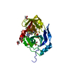 7mm2C  7mm3C 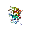 7mm4C 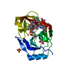 7mm5C 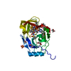 7mm6C  7mm7C  7mm9C  7mmaC  7mmbC  7mmcC  7mmdC 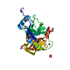 7mmfC  7mmgC 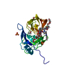 7mmhC 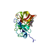 7mmiC  7mmjC 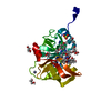 7mmkC  7mmlC  5vojS S: Starting model for refinement C: citing same article ( |
|---|---|
| Similar structure data |
-Links
 PDBj
PDBj
- Assembly
| Deposited unit | 
| ||||||||||||
|---|---|---|---|---|---|---|---|---|---|---|---|---|---|
| 1 |
| ||||||||||||
| Unit cell |
|
-Components
-Protein , 1 types, 1 molecules A
| #1: Protein | Mass: 21349.162 Da / Num. of mol.: 1 Source method: isolated from a genetically manipulated source Source: (gene. exp.) Hepacivirus C / Production host:  |
|---|
-Non-polymers , 5 types, 235 molecules 


| #2: Chemical | ChemComp-ZN /  Mass: 65.409 Da / Num. of mol.: 1 / Source method: obtained synthetically / Formula: Zn Mass: 65.409 Da / Num. of mol.: 1 / Source method: obtained synthetically / Formula: Zn | ||||
|---|---|---|---|---|---|
| #3: Chemical | ChemComp-ZJM / ( Mass: 784.894 Da / Num. of mol.: 1 / Source method: obtained synthetically / Formula: C38H49FN6O9S Mass: 784.894 Da / Num. of mol.: 1 / Source method: obtained synthetically / Formula: C38H49FN6O9S | ||||
| #4: Chemical | ChemComp-EDO /  Mass: 62.068 Da / Num. of mol.: 10 / Source method: obtained synthetically / Formula: C2H6O2 Mass: 62.068 Da / Num. of mol.: 10 / Source method: obtained synthetically / Formula: C2H6O2#5: Chemical | ChemComp-SO4 / |  Mass: 96.063 Da / Num. of mol.: 1 / Source method: obtained synthetically / Formula: SO4 Mass: 96.063 Da / Num. of mol.: 1 / Source method: obtained synthetically / Formula: SO4#6: Water | ChemComp-HOH / | Mass: 18.015 Da / Num. of mol.: 222 / Source method: isolated from a natural source / Formula: H2O |
-Details
| Has ligand of interest | Y |
|---|
-Experimental details
-Experiment
| Experiment | Method: X-RAY DIFFRACTION / Number of used crystals: 1 |
|---|
- Sample preparation
| Crystal | Density Matthews: 3.04 Å3/Da / Density % sol: 59.6 % |
|---|---|
| Crystal grow | Temperature: 298 K / Method: vapor diffusion, hanging drop / pH: 6.5 Details: 100 mM MES, pH 6.5, 4% w/v ammonium sulfate, 20-26% PEG3350, cryoprotectant = same + 15% ethylene glycol |
-Data collection
| Diffraction | Mean temperature: 100 K / Serial crystal experiment: N |
|---|---|
| Diffraction source | Source: ROTATING ANODE / Type: RIGAKU MICROMAX-007 HF / Wavelength: 1.54178 Å |
| Detector | Type: RIGAKU SATURN 944 / Detector: CCD / Date: Jun 24, 2019 |
| Radiation | Protocol: SINGLE WAVELENGTH / Monochromatic (M) / Laue (L): M / Scattering type: x-ray |
| Radiation wavelength | Wavelength: 1.54178 Å / Relative weight: 1 |
| Reflection | Resolution: 1.43→26.28 Å / Num. obs: 47936 / % possible obs: 97.74 % / Redundancy: 3.7 % / Biso Wilson estimate: 15.5 Å2 / CC1/2: 0.973 / Net I/σ(I): 16.57 |
| Reflection shell | Resolution: 1.43→1.481 Å / Num. unique obs: 4388 / CC1/2: 0.88 |
- Processing
| Software |
| |||||||||||||||||||||||||||||||||||||||||||||||||||||||||||||||||||||||||||||||||||||||||||||||||||||||||
|---|---|---|---|---|---|---|---|---|---|---|---|---|---|---|---|---|---|---|---|---|---|---|---|---|---|---|---|---|---|---|---|---|---|---|---|---|---|---|---|---|---|---|---|---|---|---|---|---|---|---|---|---|---|---|---|---|---|---|---|---|---|---|---|---|---|---|---|---|---|---|---|---|---|---|---|---|---|---|---|---|---|---|---|---|---|---|---|---|---|---|---|---|---|---|---|---|---|---|---|---|---|---|---|---|---|---|
| Refinement | Method to determine structure: MOLECULAR REPLACEMENT Starting model: PDB entry 5VOJ Resolution: 1.43→26.28 Å / SU ML: 0.1171 / Cross valid method: FREE R-VALUE / σ(F): 1.34 / Phase error: 19.7368 Stereochemistry target values: GeoStd + Monomer Library + CDL v1.2
| |||||||||||||||||||||||||||||||||||||||||||||||||||||||||||||||||||||||||||||||||||||||||||||||||||||||||
| Solvent computation | Shrinkage radii: 0.9 Å / VDW probe radii: 1.11 Å / Solvent model: FLAT BULK SOLVENT MODEL | |||||||||||||||||||||||||||||||||||||||||||||||||||||||||||||||||||||||||||||||||||||||||||||||||||||||||
| Displacement parameters | Biso mean: 21.06 Å2 | |||||||||||||||||||||||||||||||||||||||||||||||||||||||||||||||||||||||||||||||||||||||||||||||||||||||||
| Refinement step | Cycle: LAST / Resolution: 1.43→26.28 Å
| |||||||||||||||||||||||||||||||||||||||||||||||||||||||||||||||||||||||||||||||||||||||||||||||||||||||||
| Refine LS restraints |
| |||||||||||||||||||||||||||||||||||||||||||||||||||||||||||||||||||||||||||||||||||||||||||||||||||||||||
| LS refinement shell |
|
