 Movie
Movie Controller


+ Open data

- Basic information
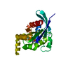
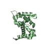
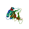
| Entry | Database: PDB / ID: 7mld | ||||||
|---|---|---|---|---|---|---|---|
| Title | PYL10 bound to the ABA pan-antagonist antabactin | ||||||
 Components Components | Abscisic acid receptor PYL10 | ||||||
 Keywords Keywords | PLANT PROTEIN/ANTAGONIST / PYR/PYL/RCAR / PYL10 / HORMONE RECEPTOR / PLANT PROTEIN / PLANT PROTEIN-ANTAGONIST complex | ||||||
| Function / homology |  Function and homology information Function and homology informationabscisic acid binding / abscisic acid-activated signaling pathway / protein phosphatase inhibitor activity / signaling receptor activity / protein homodimerization activity / nucleus / plasma membrane / cytoplasm Similarity search - Function | ||||||
| Biological species |  | ||||||
| Method |  X-RAY DIFFRACTION / MOLECULAR REPLACEMENT / Resolution: 1.8 Å X-RAY DIFFRACTION / MOLECULAR REPLACEMENT / Resolution: 1.8 Å | ||||||
 Authors Authors | Peterson, F.C. / Vaidya, A.S. / Volkman, B.F. / Cutler, S.R. | ||||||
| Funding support |  United States, 1items United States, 1items
| ||||||
 Citation Citation | Journal: Proc.Natl.Acad.Sci.USA / Year: 2021 Title: Click-to-lead design of a picomolar ABA receptor antagonist with potent activity in vivo. Authors: Vaidya, A.S. / Peterson, F.C. / Eckhardt, J. / Xing, Z. / Park, S.Y. / Dejonghe, W. / Takeuchi, J. / Pri-Tal, O. / Faria, J. / Elzinga, D. / Volkman, B.F. / Todoroki, Y. / Mosquna, A. / ...Authors: Vaidya, A.S. / Peterson, F.C. / Eckhardt, J. / Xing, Z. / Park, S.Y. / Dejonghe, W. / Takeuchi, J. / Pri-Tal, O. / Faria, J. / Elzinga, D. / Volkman, B.F. / Todoroki, Y. / Mosquna, A. / Okamoto, M. / Cutler, S.R. | ||||||
| History |
|
- Structure visualization
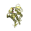
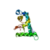
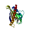
| Structure viewer | Molecule: MolmilJmol/JSmol |
|---|
- Downloads & links
-Download
| PDBx/mmCIF format | 7mld.cif.gz | 61.8 KB | Display | PDBx/mmCIF format |
|---|---|---|---|---|
| PDB format | pdb7mld.ent.gz | 34.7 KB | Display | PDB format |
| PDBx/mmJSON format | 7mld.json.gz | Tree view | PDBx/mmJSON format | |
| Others |  Other downloads Other downloads |
-Validation report
| Summary document | 7mld_validation.pdf.gz | 723.9 KB | Display | wwPDB validaton report |
|---|---|---|---|---|
| Full document | 7mld_full_validation.pdf.gz | 725.1 KB | Display | |
| Data in XML | 7mld_validation.xml.gz | 10.8 KB | Display | |
| Data in CIF | 7mld_validation.cif.gz | 15.2 KB | Display | |
| Arichive directory | https://data.pdbj.org/pub/pdb/validation_reports/ml/7mldftp://data.pdbj.org/pub/pdb/validation_reports/ml/7mld | HTTPS FTP |
-Related structure data
| Related structure data | 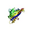 7mlcC 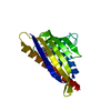 6nwbS S: Starting model for refinement C: citing same article ( |
|---|---|
| Similar structure data |


-Links
 PDBj
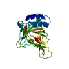
PDBj- Assembly
| Deposited unit | 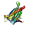
| ||||||||||||
|---|---|---|---|---|---|---|---|---|---|---|---|---|---|
| 1 |
| ||||||||||||
| Unit cell |
| ||||||||||||
| Components on special symmetry positions |
|
-Components
| #1: Protein | Mass: 17747.307 Da / Num. of mol.: 1 Source method: isolated from a genetically manipulated source Source: (gene. exp.)  |
|---|---|
| #2: Chemical | ChemComp-ZLG / 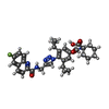  Mass: 610.678 Da / Num. of mol.: 1 / Source method: obtained synthetically / Formula: C34H35FN6O4 / Feature type: SUBJECT OF INVESTIGATION Mass: 610.678 Da / Num. of mol.: 1 / Source method: obtained synthetically / Formula: C34H35FN6O4 / Feature type: SUBJECT OF INVESTIGATION |
| #3: Chemical | ChemComp-CIT /   Mass: 192.124 Da / Num. of mol.: 1 / Source method: obtained synthetically / Formula: C6H8O7 Mass: 192.124 Da / Num. of mol.: 1 / Source method: obtained synthetically / Formula: C6H8O7 |
| #4: Water | ChemComp-HOH /  Mass: 18.015 Da / Num. of mol.: 201 / Source method: isolated from a natural source / Formula: H2O Mass: 18.015 Da / Num. of mol.: 201 / Source method: isolated from a natural source / Formula: H2O |
| Has ligand of interest | Y |
| Has protein modification | Y |
-Experimental details
-Experiment
| Experiment | Method: X-RAY DIFFRACTION / Number of used crystals: 1 |
|---|
- Sample preparation
| Crystal | Density Matthews: 2.28 Å3/Da / Density % sol: 46.11 % |
|---|---|
| Crystal grow | Temperature: 292 K / Method: vapor diffusion, hanging drop / pH: 7 Details: 200 mM tribasic ammonium citrate and 22% (w/v) PEG 3350 |
-Data collection
| Diffraction | Mean temperature: 100 K / Serial crystal experiment: N |
|---|---|
| Diffraction source | Source: ROTATING ANODE / Type: RIGAKU MICROMAX-007 / Wavelength: 1.54178 Å |
| Detector | Type: RIGAKU RAXIS IV++ / Detector: IMAGE PLATE / Date: Jul 1, 2019 |
| Radiation | Protocol: SINGLE WAVELENGTH / Monochromatic (M) / Laue (L): M / Scattering type: x-ray |
| Radiation wavelength | Wavelength: 1.54178 Å / Relative weight: 1 |
| Reflection | Resolution: 1.8→50 Å / Num. obs: 14845 / % possible obs: 97 % / Redundancy: 7.1 % / Biso Wilson estimate: 19.37 Å2 / CC1/2: 1 / CC star: 1 / Rmerge(I) obs: 0.051 / Rpim(I) all: 0.02 / Rrim(I) all: 0.055 / Net I/σ(I): 25.3 |
| Reflection shell | Resolution: 1.8→1.86 Å / Rmerge(I) obs: 0.373 / Mean I/σ(I) obs: 2.6 / Num. unique obs: 1429 / CC1/2: 0.912 / CC star: 0.977 / Rpim(I) all: 0.181 / Rrim(I) all: 0.416 / % possible all: 86.8 |
- Processing
| Software |
| |||||||||||||||||||||||||||||||||||||||||||||||||||||||||||||||||||||||||||||
|---|---|---|---|---|---|---|---|---|---|---|---|---|---|---|---|---|---|---|---|---|---|---|---|---|---|---|---|---|---|---|---|---|---|---|---|---|---|---|---|---|---|---|---|---|---|---|---|---|---|---|---|---|---|---|---|---|---|---|---|---|---|---|---|---|---|---|---|---|---|---|---|---|---|---|---|---|---|---|
| Refinement | Method to determine structure: MOLECULAR REPLACEMENT Starting model: 6NWB Resolution: 1.8→42.45 Å / SU ML: 0.2245 / Cross valid method: FREE R-VALUE / σ(F): 0 / Phase error: 19.9493 Stereochemistry target values: GeoStd + Monomer Library + CDL v1.2
| |||||||||||||||||||||||||||||||||||||||||||||||||||||||||||||||||||||||||||||
| Solvent computation | Shrinkage radii: 0.9 Å / VDW probe radii: 1.11 Å / Solvent model: FLAT BULK SOLVENT MODEL | |||||||||||||||||||||||||||||||||||||||||||||||||||||||||||||||||||||||||||||
| Displacement parameters | Biso mean: 21.38 Å2 | |||||||||||||||||||||||||||||||||||||||||||||||||||||||||||||||||||||||||||||
| Refinement step | Cycle: LAST / Resolution: 1.8→42.45 Å
| |||||||||||||||||||||||||||||||||||||||||||||||||||||||||||||||||||||||||||||
| Refine LS restraints |
| |||||||||||||||||||||||||||||||||||||||||||||||||||||||||||||||||||||||||||||
| LS refinement shell |
|
